Doaa Maamoun Ashour, Nada Abdel Salam Abdel Aziz, Khadiga Eltonbary, Noha Abdul Khaliq, Yasmeen Abdelaziz Fereig
{"title":"儿童双侧眼内肿物首次表现为弥散性结核:病例报告。","authors":"Doaa Maamoun Ashour, Nada Abdel Salam Abdel Aziz, Khadiga Eltonbary, Noha Abdul Khaliq, Yasmeen Abdelaziz Fereig","doi":"10.1177/25158414251356373","DOIUrl":null,"url":null,"abstract":"<p><p>An 8-year-old girl presented with left leukocoria, prompting an evaluation that revealed bilateral intraocular masses, including a right upper nasal choroidal lesion and a large left heterogeneous mass with exudative detachment. Imaging and systemic assessment uncovered multiple intracranial lesions, miliary lung lesion, a facial lesion, and a right tibial lesion. A strongly positive tuberculin test and a history of close contact with a tuberculosis (TB) patient led to the diagnosis of disseminated TB. Treatment with anti-tuberculosis therapy and systemic steroids initially resulted in improvement, including regression of the ocular tuberculomas and enhanced visual acuity. However, the patient developed severe headache due to non-communicating obstructive hydrocephalus, necessitating surgical intervention. Despite intensive care, the patient ultimately succumbed to the condition. This case highlights that ocular TB lesions can mimic intraocular tumors and underscores the importance of comprehensive evaluation, including multimodal imaging and systemic work-up, for early diagnosis and management of disseminated TB.</p>","PeriodicalId":23054,"journal":{"name":"Therapeutic Advances in Ophthalmology","volume":"17 ","pages":"25158414251356373"},"PeriodicalIF":2.3000,"publicationDate":"2025-07-13","publicationTypes":"Journal Article","fieldsOfStudy":null,"isOpenAccess":false,"openAccessPdf":"https://www.ncbi.nlm.nih.gov/pmc/articles/PMC12256744/pdf/","citationCount":"0","resultStr":"{\"title\":\"Bilateral intraocular masses in a child as a first presentation of disseminated tuberculosis: case report.\",\"authors\":\"Doaa Maamoun Ashour, Nada Abdel Salam Abdel Aziz, Khadiga Eltonbary, Noha Abdul Khaliq, Yasmeen Abdelaziz Fereig\",\"doi\":\"10.1177/25158414251356373\",\"DOIUrl\":null,\"url\":null,\"abstract\":\"<p><p>An 8-year-old girl presented with left leukocoria, prompting an evaluation that revealed bilateral intraocular masses, including a right upper nasal choroidal lesion and a large left heterogeneous mass with exudative detachment. Imaging and systemic assessment uncovered multiple intracranial lesions, miliary lung lesion, a facial lesion, and a right tibial lesion. A strongly positive tuberculin test and a history of close contact with a tuberculosis (TB) patient led to the diagnosis of disseminated TB. Treatment with anti-tuberculosis therapy and systemic steroids initially resulted in improvement, including regression of the ocular tuberculomas and enhanced visual acuity. However, the patient developed severe headache due to non-communicating obstructive hydrocephalus, necessitating surgical intervention. Despite intensive care, the patient ultimately succumbed to the condition. This case highlights that ocular TB lesions can mimic intraocular tumors and underscores the importance of comprehensive evaluation, including multimodal imaging and systemic work-up, for early diagnosis and management of disseminated TB.</p>\",\"PeriodicalId\":23054,\"journal\":{\"name\":\"Therapeutic Advances in Ophthalmology\",\"volume\":\"17 \",\"pages\":\"25158414251356373\"},\"PeriodicalIF\":2.3000,\"publicationDate\":\"2025-07-13\",\"publicationTypes\":\"Journal Article\",\"fieldsOfStudy\":null,\"isOpenAccess\":false,\"openAccessPdf\":\"https://www.ncbi.nlm.nih.gov/pmc/articles/PMC12256744/pdf/\",\"citationCount\":\"0\",\"resultStr\":null,\"platform\":\"Semanticscholar\",\"paperid\":null,\"PeriodicalName\":\"Therapeutic Advances in Ophthalmology\",\"FirstCategoryId\":\"1085\",\"ListUrlMain\":\"https://doi.org/10.1177/25158414251356373\",\"RegionNum\":0,\"RegionCategory\":null,\"ArticlePicture\":[],\"TitleCN\":null,\"AbstractTextCN\":null,\"PMCID\":null,\"EPubDate\":\"2025/1/1 0:00:00\",\"PubModel\":\"eCollection\",\"JCR\":\"Q2\",\"JCRName\":\"OPHTHALMOLOGY\",\"Score\":null,\"Total\":0}","platform":"Semanticscholar","paperid":null,"PeriodicalName":"Therapeutic Advances in Ophthalmology","FirstCategoryId":"1085","ListUrlMain":"https://doi.org/10.1177/25158414251356373","RegionNum":0,"RegionCategory":null,"ArticlePicture":[],"TitleCN":null,"AbstractTextCN":null,"PMCID":null,"EPubDate":"2025/1/1 0:00:00","PubModel":"eCollection","JCR":"Q2","JCRName":"OPHTHALMOLOGY","Score":null,"Total":0}
Bilateral intraocular masses in a child as a first presentation of disseminated tuberculosis: case report.
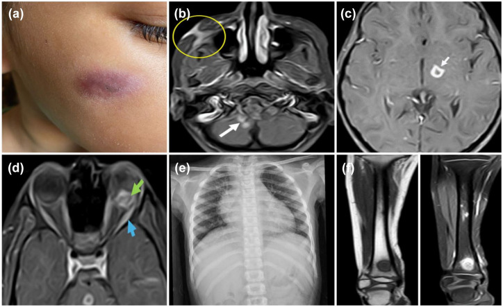
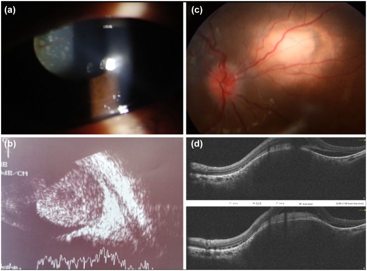
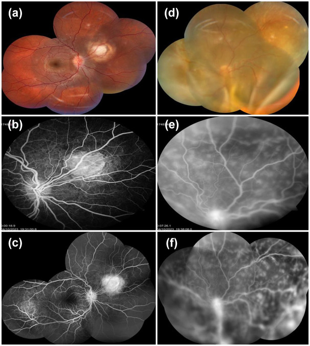
An 8-year-old girl presented with left leukocoria, prompting an evaluation that revealed bilateral intraocular masses, including a right upper nasal choroidal lesion and a large left heterogeneous mass with exudative detachment. Imaging and systemic assessment uncovered multiple intracranial lesions, miliary lung lesion, a facial lesion, and a right tibial lesion. A strongly positive tuberculin test and a history of close contact with a tuberculosis (TB) patient led to the diagnosis of disseminated TB. Treatment with anti-tuberculosis therapy and systemic steroids initially resulted in improvement, including regression of the ocular tuberculomas and enhanced visual acuity. However, the patient developed severe headache due to non-communicating obstructive hydrocephalus, necessitating surgical intervention. Despite intensive care, the patient ultimately succumbed to the condition. This case highlights that ocular TB lesions can mimic intraocular tumors and underscores the importance of comprehensive evaluation, including multimodal imaging and systemic work-up, for early diagnosis and management of disseminated TB.

 求助内容:
求助内容: 应助结果提醒方式:


