{"title":"应用合成MRI评估系统性红斑狼疮灰质结构改变。","authors":"Kemei Deng, Chengli Wu, Yuhong Qin, Wei Cui, Jing Wen, Muliang Jiang, Liling Long, Bihong T Chen","doi":"10.1136/lupus-2025-001505","DOIUrl":null,"url":null,"abstract":"<p><strong>Objectives: </strong>To assess brain grey matter alterations in patients with SLE and their correlation with neuropsychological testing using synthetic MRI (SyMRI).</p><p><strong>Methods: </strong>This prospective study enrolled patients with SLE and age, gender and education-matched healthy controls (HC). Study assessments included brain MRI using SyMRI and neuropsychological tests: Mini-Mental State Examination (MMSE), Montreal Cognitive Assessment (MoCA), Digit Span Test, Self-Rating Anxiety Scale and Self-Rating Depression Scale (SDS). SyMRI post-processing and Automated Anatomical Labeling were used for grey matter mapping. Correlation analysis was performed to assess the relationship between brain grey matter structural alterations and neuropsychological testing.</p><p><strong>Results: </strong>77 patients with SLE (57 non-neuropsychiatric SLE (non-NPSLE), 20 NPSLE) and 29 HC participants were enrolled. Patients with SLE showed reduced grey matter volume compared with HC (p<0.05). The NPSLE group exhibited more extensive increases in longitudinal (T1) and transverse (T2) relaxation times in grey matter than the non-NPSLE group (p<0.001). Proton density values were lower in patients with SLE (p<0.001). Lower brain parenchymal volume correlated with higher SLE Disease Activity Index (p<0.05). Lower MMSE/MoCA scores correlated with increased T1/T2 in the left medial cingulate and paracingulate gyri (p<0.05). Higher SDS scores correlated with increased T1/T2 in the left calcarine fissure and surrounding cortex (p<0.05). These changes were also linked to disease markers (C3, C4, immunoglobulin M, erythrocyte sedimentation rate) (p<0.05).</p><p><strong>Conclusions: </strong>Grey matter alterations in patients with SLE correlate with cognitive impairment, depression and disease activity.</p>","PeriodicalId":18126,"journal":{"name":"Lupus Science & Medicine","volume":"12 2","pages":""},"PeriodicalIF":3.5000,"publicationDate":"2025-07-13","publicationTypes":"Journal Article","fieldsOfStudy":null,"isOpenAccess":false,"openAccessPdf":"https://www.ncbi.nlm.nih.gov/pmc/articles/PMC12273088/pdf/","citationCount":"0","resultStr":"{\"title\":\"Assessing grey matter structural alterations in systemic lupus erythematosus using synthetic MRI.\",\"authors\":\"Kemei Deng, Chengli Wu, Yuhong Qin, Wei Cui, Jing Wen, Muliang Jiang, Liling Long, Bihong T Chen\",\"doi\":\"10.1136/lupus-2025-001505\",\"DOIUrl\":null,\"url\":null,\"abstract\":\"<p><strong>Objectives: </strong>To assess brain grey matter alterations in patients with SLE and their correlation with neuropsychological testing using synthetic MRI (SyMRI).</p><p><strong>Methods: </strong>This prospective study enrolled patients with SLE and age, gender and education-matched healthy controls (HC). Study assessments included brain MRI using SyMRI and neuropsychological tests: Mini-Mental State Examination (MMSE), Montreal Cognitive Assessment (MoCA), Digit Span Test, Self-Rating Anxiety Scale and Self-Rating Depression Scale (SDS). SyMRI post-processing and Automated Anatomical Labeling were used for grey matter mapping. Correlation analysis was performed to assess the relationship between brain grey matter structural alterations and neuropsychological testing.</p><p><strong>Results: </strong>77 patients with SLE (57 non-neuropsychiatric SLE (non-NPSLE), 20 NPSLE) and 29 HC participants were enrolled. Patients with SLE showed reduced grey matter volume compared with HC (p<0.05). The NPSLE group exhibited more extensive increases in longitudinal (T1) and transverse (T2) relaxation times in grey matter than the non-NPSLE group (p<0.001). Proton density values were lower in patients with SLE (p<0.001). Lower brain parenchymal volume correlated with higher SLE Disease Activity Index (p<0.05). Lower MMSE/MoCA scores correlated with increased T1/T2 in the left medial cingulate and paracingulate gyri (p<0.05). Higher SDS scores correlated with increased T1/T2 in the left calcarine fissure and surrounding cortex (p<0.05). These changes were also linked to disease markers (C3, C4, immunoglobulin M, erythrocyte sedimentation rate) (p<0.05).</p><p><strong>Conclusions: </strong>Grey matter alterations in patients with SLE correlate with cognitive impairment, depression and disease activity.</p>\",\"PeriodicalId\":18126,\"journal\":{\"name\":\"Lupus Science & Medicine\",\"volume\":\"12 2\",\"pages\":\"\"},\"PeriodicalIF\":3.5000,\"publicationDate\":\"2025-07-13\",\"publicationTypes\":\"Journal Article\",\"fieldsOfStudy\":null,\"isOpenAccess\":false,\"openAccessPdf\":\"https://www.ncbi.nlm.nih.gov/pmc/articles/PMC12273088/pdf/\",\"citationCount\":\"0\",\"resultStr\":null,\"platform\":\"Semanticscholar\",\"paperid\":null,\"PeriodicalName\":\"Lupus Science & Medicine\",\"FirstCategoryId\":\"3\",\"ListUrlMain\":\"https://doi.org/10.1136/lupus-2025-001505\",\"RegionNum\":2,\"RegionCategory\":\"医学\",\"ArticlePicture\":[],\"TitleCN\":null,\"AbstractTextCN\":null,\"PMCID\":null,\"EPubDate\":\"\",\"PubModel\":\"\",\"JCR\":\"Q1\",\"JCRName\":\"RHEUMATOLOGY\",\"Score\":null,\"Total\":0}","platform":"Semanticscholar","paperid":null,"PeriodicalName":"Lupus Science & Medicine","FirstCategoryId":"3","ListUrlMain":"https://doi.org/10.1136/lupus-2025-001505","RegionNum":2,"RegionCategory":"医学","ArticlePicture":[],"TitleCN":null,"AbstractTextCN":null,"PMCID":null,"EPubDate":"","PubModel":"","JCR":"Q1","JCRName":"RHEUMATOLOGY","Score":null,"Total":0}
Assessing grey matter structural alterations in systemic lupus erythematosus using synthetic MRI.
Objectives: To assess brain grey matter alterations in patients with SLE and their correlation with neuropsychological testing using synthetic MRI (SyMRI).
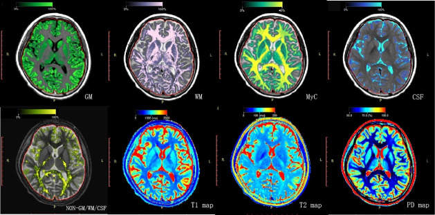
Methods: This prospective study enrolled patients with SLE and age, gender and education-matched healthy controls (HC). Study assessments included brain MRI using SyMRI and neuropsychological tests: Mini-Mental State Examination (MMSE), Montreal Cognitive Assessment (MoCA), Digit Span Test, Self-Rating Anxiety Scale and Self-Rating Depression Scale (SDS). SyMRI post-processing and Automated Anatomical Labeling were used for grey matter mapping. Correlation analysis was performed to assess the relationship between brain grey matter structural alterations and neuropsychological testing.
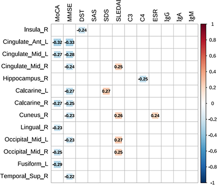
Results: 77 patients with SLE (57 non-neuropsychiatric SLE (non-NPSLE), 20 NPSLE) and 29 HC participants were enrolled. Patients with SLE showed reduced grey matter volume compared with HC (p<0.05). The NPSLE group exhibited more extensive increases in longitudinal (T1) and transverse (T2) relaxation times in grey matter than the non-NPSLE group (p<0.001). Proton density values were lower in patients with SLE (p<0.001). Lower brain parenchymal volume correlated with higher SLE Disease Activity Index (p<0.05). Lower MMSE/MoCA scores correlated with increased T1/T2 in the left medial cingulate and paracingulate gyri (p<0.05). Higher SDS scores correlated with increased T1/T2 in the left calcarine fissure and surrounding cortex (p<0.05). These changes were also linked to disease markers (C3, C4, immunoglobulin M, erythrocyte sedimentation rate) (p<0.05).
Conclusions: Grey matter alterations in patients with SLE correlate with cognitive impairment, depression and disease activity.
期刊介绍:
Lupus Science & Medicine is a global, peer reviewed, open access online journal that provides a central point for publication of basic, clinical, translational, and epidemiological studies of all aspects of lupus and related diseases. It is the first lupus-specific open access journal in the world and was developed in response to the need for a barrier-free forum for publication of groundbreaking studies in lupus. The journal publishes research on lupus from fields including, but not limited to: rheumatology, dermatology, nephrology, immunology, pediatrics, cardiology, hepatology, pulmonology, obstetrics and gynecology, and psychiatry.
 求助内容:
求助内容: 应助结果提醒方式:


