{"title":"印度尼西亚苏拉威西中部纳普河谷的脑血吸虫病。","authors":"Prawesty Diah Utami, Yunita Surya Pratiwi, Retno Budiarti, Wienta Diarsvitri","doi":"10.1155/carm/4781807","DOIUrl":null,"url":null,"abstract":"<p><p>Schistosomiasis is one of the neglected tropical diseases caused by parasitic worm infections of the genus <i>Schistosoma</i>. Involvement of the brain in schistosomiasis represents a particularly severe manifestation of the infection. Accurate diagnosis and appropriate treatment of cerebral schistosomiasis are essential, especially in our healthcare facility located in a remote area of Indonesia, where available resources are highly limited. We reported a 31-year-old female patient complaining of tonic-clonic convulsions. Before experiencing seizures, the patient reported experiencing headaches for 6 months. The patient's anamnesis regarding her social life revealed that she has been employed on a plantation for 15 years; the plantation serves as a natural habitat for snails, intermediate hosts for <i>Schistosoma</i> sp. Serological examinations were not performed due to the constraints of diagnostic instruments in the region. Cerebral schistosomiasis diagnosis was verified based on biopsy, stool examination, and CT scan results. She was admitted with a combination of steroids and praziquantel at a dosage of 60 mg/kg single dose. She was released after 14 days in satisfactory overall health. The follow-up CT scan revealed improvement, corroborated by the patient's clinical recovery. This report emphasizes the diagnostic obstacles associated with cerebral schistosomiasis, particularly in remote regions and resource-limited settings in Indonesia. Despite the absence of serological testing, a definitive diagnosis was successfully established through radiological imaging, stool microscopic examination, and brain tissue biopsy (histopathological analysis) which revealed <i>Schistosoma</i> eggs surrounded by granulomatous inflammation. The patient presented with space-occupying brain lesions and neurological symptoms, but without hepatic involvement, making the diagnosis less straightforward. This case illustrates the significance of recognizing cerebral schistosomiasis as a differential diagnosis in patients presenting cerebral lesions in endemic locations. Diagnosis of cerebral schistosomiasis based on a detailed social occupational history correlated with radiological imaging, stool microscopic examination, and brain tissue biopsy (histopathological analysis) is essential when other diagnostic tools (serological testing) are unavailable.</p>","PeriodicalId":9627,"journal":{"name":"Case Reports in Medicine","volume":"2025 ","pages":"4781807"},"PeriodicalIF":0.7000,"publicationDate":"2025-07-01","publicationTypes":"Journal Article","fieldsOfStudy":null,"isOpenAccess":false,"openAccessPdf":"https://www.ncbi.nlm.nih.gov/pmc/articles/PMC12256173/pdf/","citationCount":"0","resultStr":"{\"title\":\"Cerebral Schistosomiasis in the Napu Valley, Central Sulawesi, Indonesia.\",\"authors\":\"Prawesty Diah Utami, Yunita Surya Pratiwi, Retno Budiarti, Wienta Diarsvitri\",\"doi\":\"10.1155/carm/4781807\",\"DOIUrl\":null,\"url\":null,\"abstract\":\"<p><p>Schistosomiasis is one of the neglected tropical diseases caused by parasitic worm infections of the genus <i>Schistosoma</i>. Involvement of the brain in schistosomiasis represents a particularly severe manifestation of the infection. Accurate diagnosis and appropriate treatment of cerebral schistosomiasis are essential, especially in our healthcare facility located in a remote area of Indonesia, where available resources are highly limited. We reported a 31-year-old female patient complaining of tonic-clonic convulsions. Before experiencing seizures, the patient reported experiencing headaches for 6 months. The patient's anamnesis regarding her social life revealed that she has been employed on a plantation for 15 years; the plantation serves as a natural habitat for snails, intermediate hosts for <i>Schistosoma</i> sp. Serological examinations were not performed due to the constraints of diagnostic instruments in the region. Cerebral schistosomiasis diagnosis was verified based on biopsy, stool examination, and CT scan results. She was admitted with a combination of steroids and praziquantel at a dosage of 60 mg/kg single dose. She was released after 14 days in satisfactory overall health. The follow-up CT scan revealed improvement, corroborated by the patient's clinical recovery. This report emphasizes the diagnostic obstacles associated with cerebral schistosomiasis, particularly in remote regions and resource-limited settings in Indonesia. Despite the absence of serological testing, a definitive diagnosis was successfully established through radiological imaging, stool microscopic examination, and brain tissue biopsy (histopathological analysis) which revealed <i>Schistosoma</i> eggs surrounded by granulomatous inflammation. The patient presented with space-occupying brain lesions and neurological symptoms, but without hepatic involvement, making the diagnosis less straightforward. This case illustrates the significance of recognizing cerebral schistosomiasis as a differential diagnosis in patients presenting cerebral lesions in endemic locations. Diagnosis of cerebral schistosomiasis based on a detailed social occupational history correlated with radiological imaging, stool microscopic examination, and brain tissue biopsy (histopathological analysis) is essential when other diagnostic tools (serological testing) are unavailable.</p>\",\"PeriodicalId\":9627,\"journal\":{\"name\":\"Case Reports in Medicine\",\"volume\":\"2025 \",\"pages\":\"4781807\"},\"PeriodicalIF\":0.7000,\"publicationDate\":\"2025-07-01\",\"publicationTypes\":\"Journal Article\",\"fieldsOfStudy\":null,\"isOpenAccess\":false,\"openAccessPdf\":\"https://www.ncbi.nlm.nih.gov/pmc/articles/PMC12256173/pdf/\",\"citationCount\":\"0\",\"resultStr\":null,\"platform\":\"Semanticscholar\",\"paperid\":null,\"PeriodicalName\":\"Case Reports in Medicine\",\"FirstCategoryId\":\"1085\",\"ListUrlMain\":\"https://doi.org/10.1155/carm/4781807\",\"RegionNum\":0,\"RegionCategory\":null,\"ArticlePicture\":[],\"TitleCN\":null,\"AbstractTextCN\":null,\"PMCID\":null,\"EPubDate\":\"2025/1/1 0:00:00\",\"PubModel\":\"eCollection\",\"JCR\":\"Q3\",\"JCRName\":\"MEDICINE, GENERAL & INTERNAL\",\"Score\":null,\"Total\":0}","platform":"Semanticscholar","paperid":null,"PeriodicalName":"Case Reports in Medicine","FirstCategoryId":"1085","ListUrlMain":"https://doi.org/10.1155/carm/4781807","RegionNum":0,"RegionCategory":null,"ArticlePicture":[],"TitleCN":null,"AbstractTextCN":null,"PMCID":null,"EPubDate":"2025/1/1 0:00:00","PubModel":"eCollection","JCR":"Q3","JCRName":"MEDICINE, GENERAL & INTERNAL","Score":null,"Total":0}
Cerebral Schistosomiasis in the Napu Valley, Central Sulawesi, Indonesia.
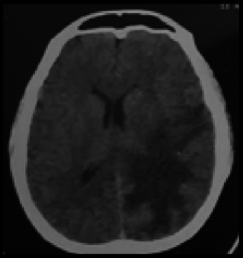
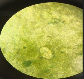
Schistosomiasis is one of the neglected tropical diseases caused by parasitic worm infections of the genus Schistosoma. Involvement of the brain in schistosomiasis represents a particularly severe manifestation of the infection. Accurate diagnosis and appropriate treatment of cerebral schistosomiasis are essential, especially in our healthcare facility located in a remote area of Indonesia, where available resources are highly limited. We reported a 31-year-old female patient complaining of tonic-clonic convulsions. Before experiencing seizures, the patient reported experiencing headaches for 6 months. The patient's anamnesis regarding her social life revealed that she has been employed on a plantation for 15 years; the plantation serves as a natural habitat for snails, intermediate hosts for Schistosoma sp. Serological examinations were not performed due to the constraints of diagnostic instruments in the region. Cerebral schistosomiasis diagnosis was verified based on biopsy, stool examination, and CT scan results. She was admitted with a combination of steroids and praziquantel at a dosage of 60 mg/kg single dose. She was released after 14 days in satisfactory overall health. The follow-up CT scan revealed improvement, corroborated by the patient's clinical recovery. This report emphasizes the diagnostic obstacles associated with cerebral schistosomiasis, particularly in remote regions and resource-limited settings in Indonesia. Despite the absence of serological testing, a definitive diagnosis was successfully established through radiological imaging, stool microscopic examination, and brain tissue biopsy (histopathological analysis) which revealed Schistosoma eggs surrounded by granulomatous inflammation. The patient presented with space-occupying brain lesions and neurological symptoms, but without hepatic involvement, making the diagnosis less straightforward. This case illustrates the significance of recognizing cerebral schistosomiasis as a differential diagnosis in patients presenting cerebral lesions in endemic locations. Diagnosis of cerebral schistosomiasis based on a detailed social occupational history correlated with radiological imaging, stool microscopic examination, and brain tissue biopsy (histopathological analysis) is essential when other diagnostic tools (serological testing) are unavailable.

 求助内容:
求助内容: 应助结果提醒方式:


