{"title":"絮状表皮植物是足癣和甲癣的病原:约2例。","authors":"Rania Besrour, Latifa Mtibaa, Faten Rabhi, Nawel Baccouchi, Abderraouf Dhaoui, Boutheina Jemli","doi":"10.11604/pamj.2025.50.102.40580","DOIUrl":null,"url":null,"abstract":"<p><p>Dermatophytia is an infection caused by keratinophilic filamentous fungi. The distribution of dermatophytes varies by country and geographic area. Epidermophyton (E) floccosum has experienced a downward trend in recent years. It is an anthropophilic dermatophyte that causes mainly skin infections in humans. We report two observations of a tinea pedis and a tinea unguium due to E. floccosum. A 70-year-old diabetic patient was admitted to dermatology to manage leg erysipelas. The patient had scaly plantar keratoderma and intertrigo of the toe-web spaces. Mycological sampling showed at direct examination mycelial filaments. A real-time Polymerase Chain reaction (PCR) allowed the identification of E. floccosum. The patient was treated with terbinafine ointment and short-term antibiotic therapy with favorable clinical course. A 70-year-old patient presented with subungual hyperkeratosis of the two big toenails, longitudinal melanonychia on the right, xanthonychia and distal onycholysis on the left. Microscopic examination of fungal colonies allowed the identification of Epidermophyton floccosum. A confirmation of the species diagnosis was carried out by real-time PCR. The patient was treated with terbinafine 250 mg daily for 6 months. Conventional techniques are, in most cases, sufficient for diagnosing superficial mycosis. However, identification is sometimes difficult, hence the importance of the contribution of molecular biology in the diagnosis.</p>","PeriodicalId":48190,"journal":{"name":"Pan African Medical Journal","volume":"50 ","pages":"102"},"PeriodicalIF":1.0000,"publicationDate":"2025-04-11","publicationTypes":"Journal Article","fieldsOfStudy":null,"isOpenAccess":false,"openAccessPdf":"https://www.ncbi.nlm.nih.gov/pmc/articles/PMC12245641/pdf/","citationCount":"0","resultStr":"{\"title\":\"<i>Epidermophyton floccosum</i>, an etiological agent of tinea pedis and tinea unguium: about two cases.\",\"authors\":\"Rania Besrour, Latifa Mtibaa, Faten Rabhi, Nawel Baccouchi, Abderraouf Dhaoui, Boutheina Jemli\",\"doi\":\"10.11604/pamj.2025.50.102.40580\",\"DOIUrl\":null,\"url\":null,\"abstract\":\"<p><p>Dermatophytia is an infection caused by keratinophilic filamentous fungi. The distribution of dermatophytes varies by country and geographic area. Epidermophyton (E) floccosum has experienced a downward trend in recent years. It is an anthropophilic dermatophyte that causes mainly skin infections in humans. We report two observations of a tinea pedis and a tinea unguium due to E. floccosum. A 70-year-old diabetic patient was admitted to dermatology to manage leg erysipelas. The patient had scaly plantar keratoderma and intertrigo of the toe-web spaces. Mycological sampling showed at direct examination mycelial filaments. A real-time Polymerase Chain reaction (PCR) allowed the identification of E. floccosum. The patient was treated with terbinafine ointment and short-term antibiotic therapy with favorable clinical course. A 70-year-old patient presented with subungual hyperkeratosis of the two big toenails, longitudinal melanonychia on the right, xanthonychia and distal onycholysis on the left. Microscopic examination of fungal colonies allowed the identification of Epidermophyton floccosum. A confirmation of the species diagnosis was carried out by real-time PCR. The patient was treated with terbinafine 250 mg daily for 6 months. Conventional techniques are, in most cases, sufficient for diagnosing superficial mycosis. However, identification is sometimes difficult, hence the importance of the contribution of molecular biology in the diagnosis.</p>\",\"PeriodicalId\":48190,\"journal\":{\"name\":\"Pan African Medical Journal\",\"volume\":\"50 \",\"pages\":\"102\"},\"PeriodicalIF\":1.0000,\"publicationDate\":\"2025-04-11\",\"publicationTypes\":\"Journal Article\",\"fieldsOfStudy\":null,\"isOpenAccess\":false,\"openAccessPdf\":\"https://www.ncbi.nlm.nih.gov/pmc/articles/PMC12245641/pdf/\",\"citationCount\":\"0\",\"resultStr\":null,\"platform\":\"Semanticscholar\",\"paperid\":null,\"PeriodicalName\":\"Pan African Medical Journal\",\"FirstCategoryId\":\"1085\",\"ListUrlMain\":\"https://doi.org/10.11604/pamj.2025.50.102.40580\",\"RegionNum\":0,\"RegionCategory\":null,\"ArticlePicture\":[],\"TitleCN\":null,\"AbstractTextCN\":null,\"PMCID\":null,\"EPubDate\":\"2025/1/1 0:00:00\",\"PubModel\":\"eCollection\",\"JCR\":\"Q4\",\"JCRName\":\"PUBLIC, ENVIRONMENTAL & OCCUPATIONAL HEALTH\",\"Score\":null,\"Total\":0}","platform":"Semanticscholar","paperid":null,"PeriodicalName":"Pan African Medical Journal","FirstCategoryId":"1085","ListUrlMain":"https://doi.org/10.11604/pamj.2025.50.102.40580","RegionNum":0,"RegionCategory":null,"ArticlePicture":[],"TitleCN":null,"AbstractTextCN":null,"PMCID":null,"EPubDate":"2025/1/1 0:00:00","PubModel":"eCollection","JCR":"Q4","JCRName":"PUBLIC, ENVIRONMENTAL & OCCUPATIONAL HEALTH","Score":null,"Total":0}
Epidermophyton floccosum, an etiological agent of tinea pedis and tinea unguium: about two cases.
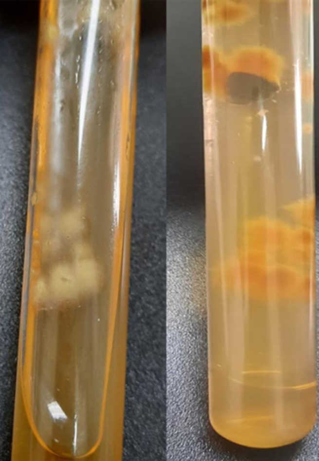
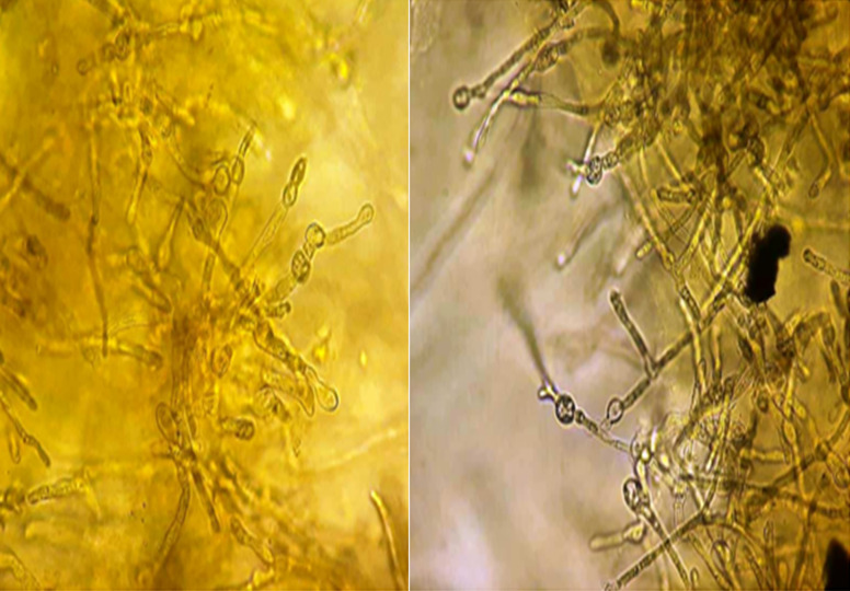
Dermatophytia is an infection caused by keratinophilic filamentous fungi. The distribution of dermatophytes varies by country and geographic area. Epidermophyton (E) floccosum has experienced a downward trend in recent years. It is an anthropophilic dermatophyte that causes mainly skin infections in humans. We report two observations of a tinea pedis and a tinea unguium due to E. floccosum. A 70-year-old diabetic patient was admitted to dermatology to manage leg erysipelas. The patient had scaly plantar keratoderma and intertrigo of the toe-web spaces. Mycological sampling showed at direct examination mycelial filaments. A real-time Polymerase Chain reaction (PCR) allowed the identification of E. floccosum. The patient was treated with terbinafine ointment and short-term antibiotic therapy with favorable clinical course. A 70-year-old patient presented with subungual hyperkeratosis of the two big toenails, longitudinal melanonychia on the right, xanthonychia and distal onycholysis on the left. Microscopic examination of fungal colonies allowed the identification of Epidermophyton floccosum. A confirmation of the species diagnosis was carried out by real-time PCR. The patient was treated with terbinafine 250 mg daily for 6 months. Conventional techniques are, in most cases, sufficient for diagnosing superficial mycosis. However, identification is sometimes difficult, hence the importance of the contribution of molecular biology in the diagnosis.

 求助内容:
求助内容: 应助结果提醒方式:


