Juraj Marton, Radovan Žižka, Rami Dabdoub, Zdeněk Pokorný
{"title":"上颌第三磨牙牙根解剖不良的半即时自体移植一例。","authors":"Juraj Marton, Radovan Žižka, Rami Dabdoub, Zdeněk Pokorný","doi":"10.1155/crid/6655016","DOIUrl":null,"url":null,"abstract":"<p><p>Tooth autotransplantation is a procedure in which a donor tooth is transplanted within the same patient's jaw to replace a missing tooth. Donor tooth root morphology and periodontal ligament integrity are key factors influencing success. We report a semi-immediate autotransplantation of a maxillary third molar with a 90° divergent root into a site previously affected by a periapical abscess. After the extraction of the compromised tooth (#15) and removal of the interradicular septum, the site was left to heal to allow soft tissue closure and infection resolution. During this period, CBCT imaging and a 3D-printed donor tooth replica (CARP model) were used to plan the procedure, including the intended root amputation. Two weeks later, autotransplantation was performed. The recipient site only required soft tissue and granulation tissue management, with no additional bone preparation, allowing for a minimally traumatic approach. The donor tooth was transplanted with an extraoral time of under 6 min. At the 18-month follow-up, the tooth remained functional, asymptomatic, and radiographically stable. This case highlights the feasibility of delayed autotransplantation following infection and the clinical value of combining imaging with prototyping in surgical planning-particularly when dealing with donor teeth with unfavorable root anatomy.</p>","PeriodicalId":46841,"journal":{"name":"Case Reports in Dentistry","volume":"2025 ","pages":"6655016"},"PeriodicalIF":0.9000,"publicationDate":"2025-07-04","publicationTypes":"Journal Article","fieldsOfStudy":null,"isOpenAccess":false,"openAccessPdf":"https://www.ncbi.nlm.nih.gov/pmc/articles/PMC12253988/pdf/","citationCount":"0","resultStr":"{\"title\":\"Semi-Immediate Autotransplantation of a Maxillary Third Molar With Unfavorable Root Anatomy: A Case Report.\",\"authors\":\"Juraj Marton, Radovan Žižka, Rami Dabdoub, Zdeněk Pokorný\",\"doi\":\"10.1155/crid/6655016\",\"DOIUrl\":null,\"url\":null,\"abstract\":\"<p><p>Tooth autotransplantation is a procedure in which a donor tooth is transplanted within the same patient's jaw to replace a missing tooth. Donor tooth root morphology and periodontal ligament integrity are key factors influencing success. We report a semi-immediate autotransplantation of a maxillary third molar with a 90° divergent root into a site previously affected by a periapical abscess. After the extraction of the compromised tooth (#15) and removal of the interradicular septum, the site was left to heal to allow soft tissue closure and infection resolution. During this period, CBCT imaging and a 3D-printed donor tooth replica (CARP model) were used to plan the procedure, including the intended root amputation. Two weeks later, autotransplantation was performed. The recipient site only required soft tissue and granulation tissue management, with no additional bone preparation, allowing for a minimally traumatic approach. The donor tooth was transplanted with an extraoral time of under 6 min. At the 18-month follow-up, the tooth remained functional, asymptomatic, and radiographically stable. This case highlights the feasibility of delayed autotransplantation following infection and the clinical value of combining imaging with prototyping in surgical planning-particularly when dealing with donor teeth with unfavorable root anatomy.</p>\",\"PeriodicalId\":46841,\"journal\":{\"name\":\"Case Reports in Dentistry\",\"volume\":\"2025 \",\"pages\":\"6655016\"},\"PeriodicalIF\":0.9000,\"publicationDate\":\"2025-07-04\",\"publicationTypes\":\"Journal Article\",\"fieldsOfStudy\":null,\"isOpenAccess\":false,\"openAccessPdf\":\"https://www.ncbi.nlm.nih.gov/pmc/articles/PMC12253988/pdf/\",\"citationCount\":\"0\",\"resultStr\":null,\"platform\":\"Semanticscholar\",\"paperid\":null,\"PeriodicalName\":\"Case Reports in Dentistry\",\"FirstCategoryId\":\"1085\",\"ListUrlMain\":\"https://doi.org/10.1155/crid/6655016\",\"RegionNum\":0,\"RegionCategory\":null,\"ArticlePicture\":[],\"TitleCN\":null,\"AbstractTextCN\":null,\"PMCID\":null,\"EPubDate\":\"2025/1/1 0:00:00\",\"PubModel\":\"eCollection\",\"JCR\":\"Q4\",\"JCRName\":\"DENTISTRY, ORAL SURGERY & MEDICINE\",\"Score\":null,\"Total\":0}","platform":"Semanticscholar","paperid":null,"PeriodicalName":"Case Reports in Dentistry","FirstCategoryId":"1085","ListUrlMain":"https://doi.org/10.1155/crid/6655016","RegionNum":0,"RegionCategory":null,"ArticlePicture":[],"TitleCN":null,"AbstractTextCN":null,"PMCID":null,"EPubDate":"2025/1/1 0:00:00","PubModel":"eCollection","JCR":"Q4","JCRName":"DENTISTRY, ORAL SURGERY & MEDICINE","Score":null,"Total":0}
Semi-Immediate Autotransplantation of a Maxillary Third Molar With Unfavorable Root Anatomy: A Case Report.
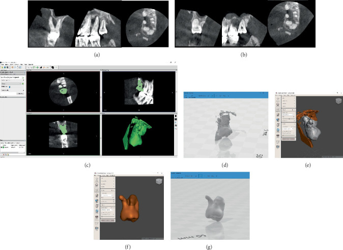
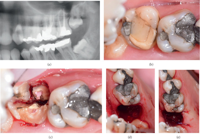
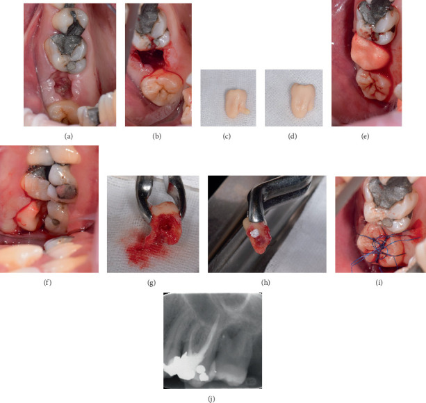
Tooth autotransplantation is a procedure in which a donor tooth is transplanted within the same patient's jaw to replace a missing tooth. Donor tooth root morphology and periodontal ligament integrity are key factors influencing success. We report a semi-immediate autotransplantation of a maxillary third molar with a 90° divergent root into a site previously affected by a periapical abscess. After the extraction of the compromised tooth (#15) and removal of the interradicular septum, the site was left to heal to allow soft tissue closure and infection resolution. During this period, CBCT imaging and a 3D-printed donor tooth replica (CARP model) were used to plan the procedure, including the intended root amputation. Two weeks later, autotransplantation was performed. The recipient site only required soft tissue and granulation tissue management, with no additional bone preparation, allowing for a minimally traumatic approach. The donor tooth was transplanted with an extraoral time of under 6 min. At the 18-month follow-up, the tooth remained functional, asymptomatic, and radiographically stable. This case highlights the feasibility of delayed autotransplantation following infection and the clinical value of combining imaging with prototyping in surgical planning-particularly when dealing with donor teeth with unfavorable root anatomy.
期刊介绍:
Case Reports in Dentistry is a peer-reviewed, Open Access journal that publishes case reports and case series in all areas of dentistry, including periodontal diseases, dental implants, oral pathology, as well as oral and maxillofacial surgery.

 求助内容:
求助内容: 应助结果提醒方式:


