人类非小细胞肺癌组织学样品的原子力显微镜表征。
IF 4.6
2区 医学
Q2 MEDICINE, RESEARCH & EXPERIMENTAL
Nanomedicine : nanotechnology, biology, and medicine
Pub Date : 2025-07-12
DOI:10.1016/j.nano.2025.102844
引用次数: 0
摘要
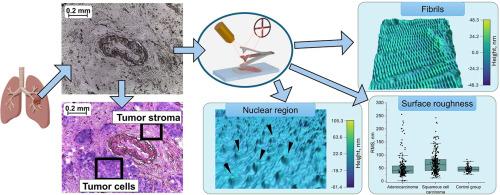
本研究利用原子力显微镜(AFM)分析了26例非小细胞肺癌组织,特别是腺癌和鳞状细胞癌的术中冷冻组织学样本。两种亚型的肿瘤细胞区域和基质之间存在明显且可测量的结构差异。核区形态在不同的细胞群中有所不同——从间质肿瘤细胞的核区模糊到非癌性支气管上皮细胞的核部分撕裂。这一观察结果可能表明核结构特征的不同。与对照样品相比,两种癌症类型的间质区域显示出更密集的条纹原纤维覆盖,胶原蛋白的周期性特征为67 nm。在切片(肿瘤和非恶性)中随机选择的部位进行标准表面粗糙度测试,表明有可能从对照组织学样本中区分恶性。结合原子力显微镜与组织病理学分析可以提供额外的结构洞察与恶性肿瘤相关的肺组织改变。本文章由计算机程序翻译,如有差异,请以英文原文为准。
Characterization of human non-small cell lung cancer histology samples by atomic force microscopy
This study utilized atomic force microscopy (AFM) to analyze intraoperative frozen histology samples from 26 patients with non-small cell lung cancer tissue, specifically adenocarcinoma and squamous cell carcinoma. Clear and measurable structural differences were identified between tumor cell regions and stroma of the two subtypes. Nuclear region morphologies varied among cell groups – ranging from indistinct nuclear regions in stromal tumor cells to partially torn nuclei in non-cancerous bronchial epithelial cells. This observation may indicate differences in nuclear structure characteristics. Compared to control samples, stromal regions of both cancer types exhibited denser coverage of striated fibrils with a 67 nm periodicity characteristic to collagen. Standard surface roughness tests on randomly selected sites within sections (tumor and non-malignant) suggest potential for distinguishing malignant from control histology samples. Combining atomic force microscopy with histopathological analysis may offer additional structural insight into lung tissue alterations associated with malignancy.
求助全文
通过发布文献求助,成功后即可免费获取论文全文。
去求助
来源期刊
CiteScore
11.10
自引率
0.00%
发文量
133
审稿时长
42 days
期刊介绍:
The mission of Nanomedicine: Nanotechnology, Biology, and Medicine (Nanomedicine: NBM) is to promote the emerging interdisciplinary field of nanomedicine.
Nanomedicine: NBM is an international, peer-reviewed journal presenting novel, significant, and interdisciplinary theoretical and experimental results related to nanoscience and nanotechnology in the life and health sciences. Content includes basic, translational, and clinical research addressing diagnosis, treatment, monitoring, prediction, and prevention of diseases.
 求助内容:
求助内容: 应助结果提醒方式:


