Yusuke Haruna, Mizuki Tagami, Gen Kinari, Atsushi Sakai, Shigeru Honda
{"title":"增殖性糖尿病视网膜病变术后玻璃体出血与术前光学相干断层血管造影评价的关系。","authors":"Yusuke Haruna, Mizuki Tagami, Gen Kinari, Atsushi Sakai, Shigeru Honda","doi":"10.1155/joph/7839246","DOIUrl":null,"url":null,"abstract":"<p><p><b>Purpose:</b> To measure and compare the extent of retinal neovascularization using optical coherence tomography angiography (OCTA) between patients with good postoperative outcomes for proliferative diabetic retinopathy (PDR) requiring surgical treatment and patients with vitreous hemorrhage (VH). <b>Methods:</b> This retrospective study included patients who were diagnosed with PDR between January 2022 and December 2023 and underwent vitreous surgery. Cases that developed postoperative VH were classified as the VH group, and cases with good postoperative progress were classified as the control group. The extent of retinal neovascularization was measured from preoperative and postoperative images of the two groups taken by OCTA measured with a widefield optical coherence tomography (Canon, Xephilio OCT-A1), and a comparative study was conducted. <b>Results:</b> The VH group consisted of 8 patients with 11 eyes (4 men and 4 women) with a mean age of 49.7 ± 14.2 years, while the control group consisted of 23 patients with 26 eyes (19 men and 4 women) with a mean age of 56.9 ± 11.8 years. The preoperative retinal neovascular area was 50,233.7 ± 38,581.1 (pixels) in the VH group and 17,155.4 ± 27,950.2 (pixels) in the control group, showing a significant difference (<i>p</i>=0.046). The postoperative retinal neovascular area was 36,315.7 ± 44,311.8 (pixels) in the VH group and 2261.0 ± 9072.2 (pixels) in the control group, showing a significant difference (<i>p</i>=0.046), but there was no significant difference in the reduction rate of the neovascular area before and after surgery (<i>p</i>=0.30). <b>Conclusions:</b> In PDR developing VH after vitrectomy surgery, the appearance of neovascularization seen on pre- and postoperative OCTA is significantly more extensive than in cases that do not develop postoperative VH, and OCTA may be useful for preoperative evaluation.</p>","PeriodicalId":16674,"journal":{"name":"Journal of Ophthalmology","volume":"2025 ","pages":"7839246"},"PeriodicalIF":1.9000,"publicationDate":"2025-07-05","publicationTypes":"Journal Article","fieldsOfStudy":null,"isOpenAccess":false,"openAccessPdf":"https://www.ncbi.nlm.nih.gov/pmc/articles/PMC12255494/pdf/","citationCount":"0","resultStr":"{\"title\":\"Correlation Between Postoperative Vitreous Hemorrhage and Preoperative Evaluation of Optical Coherence Tomography Angiography in Proliferative Diabetic Retinopathy Surgery.\",\"authors\":\"Yusuke Haruna, Mizuki Tagami, Gen Kinari, Atsushi Sakai, Shigeru Honda\",\"doi\":\"10.1155/joph/7839246\",\"DOIUrl\":null,\"url\":null,\"abstract\":\"<p><p><b>Purpose:</b> To measure and compare the extent of retinal neovascularization using optical coherence tomography angiography (OCTA) between patients with good postoperative outcomes for proliferative diabetic retinopathy (PDR) requiring surgical treatment and patients with vitreous hemorrhage (VH). <b>Methods:</b> This retrospective study included patients who were diagnosed with PDR between January 2022 and December 2023 and underwent vitreous surgery. Cases that developed postoperative VH were classified as the VH group, and cases with good postoperative progress were classified as the control group. The extent of retinal neovascularization was measured from preoperative and postoperative images of the two groups taken by OCTA measured with a widefield optical coherence tomography (Canon, Xephilio OCT-A1), and a comparative study was conducted. <b>Results:</b> The VH group consisted of 8 patients with 11 eyes (4 men and 4 women) with a mean age of 49.7 ± 14.2 years, while the control group consisted of 23 patients with 26 eyes (19 men and 4 women) with a mean age of 56.9 ± 11.8 years. The preoperative retinal neovascular area was 50,233.7 ± 38,581.1 (pixels) in the VH group and 17,155.4 ± 27,950.2 (pixels) in the control group, showing a significant difference (<i>p</i>=0.046). The postoperative retinal neovascular area was 36,315.7 ± 44,311.8 (pixels) in the VH group and 2261.0 ± 9072.2 (pixels) in the control group, showing a significant difference (<i>p</i>=0.046), but there was no significant difference in the reduction rate of the neovascular area before and after surgery (<i>p</i>=0.30). <b>Conclusions:</b> In PDR developing VH after vitrectomy surgery, the appearance of neovascularization seen on pre- and postoperative OCTA is significantly more extensive than in cases that do not develop postoperative VH, and OCTA may be useful for preoperative evaluation.</p>\",\"PeriodicalId\":16674,\"journal\":{\"name\":\"Journal of Ophthalmology\",\"volume\":\"2025 \",\"pages\":\"7839246\"},\"PeriodicalIF\":1.9000,\"publicationDate\":\"2025-07-05\",\"publicationTypes\":\"Journal Article\",\"fieldsOfStudy\":null,\"isOpenAccess\":false,\"openAccessPdf\":\"https://www.ncbi.nlm.nih.gov/pmc/articles/PMC12255494/pdf/\",\"citationCount\":\"0\",\"resultStr\":null,\"platform\":\"Semanticscholar\",\"paperid\":null,\"PeriodicalName\":\"Journal of Ophthalmology\",\"FirstCategoryId\":\"3\",\"ListUrlMain\":\"https://doi.org/10.1155/joph/7839246\",\"RegionNum\":4,\"RegionCategory\":\"医学\",\"ArticlePicture\":[],\"TitleCN\":null,\"AbstractTextCN\":null,\"PMCID\":null,\"EPubDate\":\"2025/1/1 0:00:00\",\"PubModel\":\"eCollection\",\"JCR\":\"Q3\",\"JCRName\":\"OPHTHALMOLOGY\",\"Score\":null,\"Total\":0}","platform":"Semanticscholar","paperid":null,"PeriodicalName":"Journal of Ophthalmology","FirstCategoryId":"3","ListUrlMain":"https://doi.org/10.1155/joph/7839246","RegionNum":4,"RegionCategory":"医学","ArticlePicture":[],"TitleCN":null,"AbstractTextCN":null,"PMCID":null,"EPubDate":"2025/1/1 0:00:00","PubModel":"eCollection","JCR":"Q3","JCRName":"OPHTHALMOLOGY","Score":null,"Total":0}
Correlation Between Postoperative Vitreous Hemorrhage and Preoperative Evaluation of Optical Coherence Tomography Angiography in Proliferative Diabetic Retinopathy Surgery.
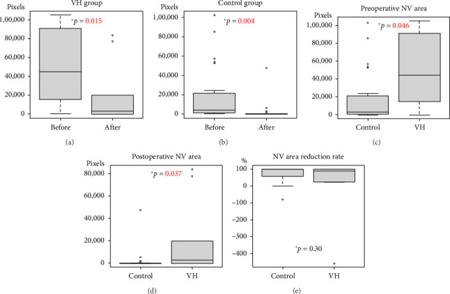
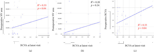
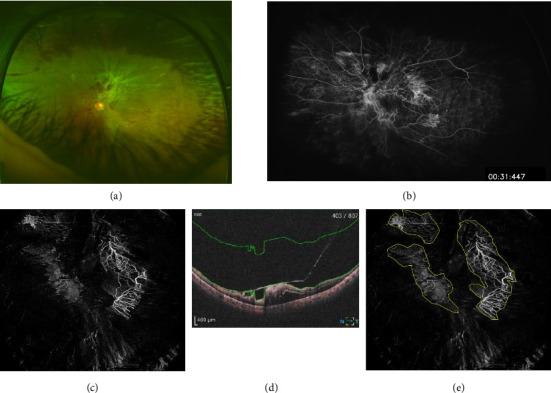
Purpose: To measure and compare the extent of retinal neovascularization using optical coherence tomography angiography (OCTA) between patients with good postoperative outcomes for proliferative diabetic retinopathy (PDR) requiring surgical treatment and patients with vitreous hemorrhage (VH). Methods: This retrospective study included patients who were diagnosed with PDR between January 2022 and December 2023 and underwent vitreous surgery. Cases that developed postoperative VH were classified as the VH group, and cases with good postoperative progress were classified as the control group. The extent of retinal neovascularization was measured from preoperative and postoperative images of the two groups taken by OCTA measured with a widefield optical coherence tomography (Canon, Xephilio OCT-A1), and a comparative study was conducted. Results: The VH group consisted of 8 patients with 11 eyes (4 men and 4 women) with a mean age of 49.7 ± 14.2 years, while the control group consisted of 23 patients with 26 eyes (19 men and 4 women) with a mean age of 56.9 ± 11.8 years. The preoperative retinal neovascular area was 50,233.7 ± 38,581.1 (pixels) in the VH group and 17,155.4 ± 27,950.2 (pixels) in the control group, showing a significant difference (p=0.046). The postoperative retinal neovascular area was 36,315.7 ± 44,311.8 (pixels) in the VH group and 2261.0 ± 9072.2 (pixels) in the control group, showing a significant difference (p=0.046), but there was no significant difference in the reduction rate of the neovascular area before and after surgery (p=0.30). Conclusions: In PDR developing VH after vitrectomy surgery, the appearance of neovascularization seen on pre- and postoperative OCTA is significantly more extensive than in cases that do not develop postoperative VH, and OCTA may be useful for preoperative evaluation.
期刊介绍:
Journal of Ophthalmology is a peer-reviewed, Open Access journal that publishes original research articles, review articles, and clinical studies related to the anatomy, physiology and diseases of the eye. Submissions should focus on new diagnostic and surgical techniques, instrument and therapy updates, as well as clinical trials and research findings.
 求助内容:
求助内容: 应助结果提醒方式:


