{"title":"Waardenburg综合征在一个家庭。","authors":"M Niveditha, Priya Prathap, Neelakandhan Asokan","doi":"10.4103/ijt.ijt_134_22","DOIUrl":null,"url":null,"abstract":"<p><p>Waardenburg syndrome (WS) is an autosomal dominant genetic disease with an estimated prevalence of 1 in 20,000-40,000. An 8-year-old boy, born to nonconsanguineous parents, presented with complaints of areas of depigmentation on the forehead and right leg since birth. On examination, there was a well-defined amelanotic macule on the center of the forehead of size 5 cm × 2.5 cm with a few spots of normal pigmentation, a forelock of white hair on the frontal area of the scalp, and a well-defined amelanotic macule of size 6 cm × 4 cm with a small central area of normal pigmentation on the posterior part of the right leg. He had scoliosis of the spine in the thoracic region. The nasal root was broad with widely separated inner canthi. There was exotropia and microcornea of the right eye with a visual acuity of 6/24. Fundus examination of the right eye showed a large disc, disc coloboma, peripapillary atrophy, and pigmentary changes in the fovea. The left eye was normal. There was no hearing defect. His father and two siblings too had patchy amelanosis in a similar distribution. They probably represent a limited expression of the same disease. All of them meet the diagnostic criteria for WS. WS is rare with only <100 cases reported worldwide.</p>","PeriodicalId":14417,"journal":{"name":"International Journal of Trichology","volume":"17 1","pages":"73-76"},"PeriodicalIF":0.0000,"publicationDate":"2025-01-01","publicationTypes":"Journal Article","fieldsOfStudy":null,"isOpenAccess":false,"openAccessPdf":"https://www.ncbi.nlm.nih.gov/pmc/articles/PMC12251972/pdf/","citationCount":"0","resultStr":"{\"title\":\"Waardenburg Syndrome in a Family.\",\"authors\":\"M Niveditha, Priya Prathap, Neelakandhan Asokan\",\"doi\":\"10.4103/ijt.ijt_134_22\",\"DOIUrl\":null,\"url\":null,\"abstract\":\"<p><p>Waardenburg syndrome (WS) is an autosomal dominant genetic disease with an estimated prevalence of 1 in 20,000-40,000. An 8-year-old boy, born to nonconsanguineous parents, presented with complaints of areas of depigmentation on the forehead and right leg since birth. On examination, there was a well-defined amelanotic macule on the center of the forehead of size 5 cm × 2.5 cm with a few spots of normal pigmentation, a forelock of white hair on the frontal area of the scalp, and a well-defined amelanotic macule of size 6 cm × 4 cm with a small central area of normal pigmentation on the posterior part of the right leg. He had scoliosis of the spine in the thoracic region. The nasal root was broad with widely separated inner canthi. There was exotropia and microcornea of the right eye with a visual acuity of 6/24. Fundus examination of the right eye showed a large disc, disc coloboma, peripapillary atrophy, and pigmentary changes in the fovea. The left eye was normal. There was no hearing defect. His father and two siblings too had patchy amelanosis in a similar distribution. They probably represent a limited expression of the same disease. All of them meet the diagnostic criteria for WS. WS is rare with only <100 cases reported worldwide.</p>\",\"PeriodicalId\":14417,\"journal\":{\"name\":\"International Journal of Trichology\",\"volume\":\"17 1\",\"pages\":\"73-76\"},\"PeriodicalIF\":0.0000,\"publicationDate\":\"2025-01-01\",\"publicationTypes\":\"Journal Article\",\"fieldsOfStudy\":null,\"isOpenAccess\":false,\"openAccessPdf\":\"https://www.ncbi.nlm.nih.gov/pmc/articles/PMC12251972/pdf/\",\"citationCount\":\"0\",\"resultStr\":null,\"platform\":\"Semanticscholar\",\"paperid\":null,\"PeriodicalName\":\"International Journal of Trichology\",\"FirstCategoryId\":\"1085\",\"ListUrlMain\":\"https://doi.org/10.4103/ijt.ijt_134_22\",\"RegionNum\":0,\"RegionCategory\":null,\"ArticlePicture\":[],\"TitleCN\":null,\"AbstractTextCN\":null,\"PMCID\":null,\"EPubDate\":\"2025/6/23 0:00:00\",\"PubModel\":\"Epub\",\"JCR\":\"Q2\",\"JCRName\":\"Medicine\",\"Score\":null,\"Total\":0}","platform":"Semanticscholar","paperid":null,"PeriodicalName":"International Journal of Trichology","FirstCategoryId":"1085","ListUrlMain":"https://doi.org/10.4103/ijt.ijt_134_22","RegionNum":0,"RegionCategory":null,"ArticlePicture":[],"TitleCN":null,"AbstractTextCN":null,"PMCID":null,"EPubDate":"2025/6/23 0:00:00","PubModel":"Epub","JCR":"Q2","JCRName":"Medicine","Score":null,"Total":0}
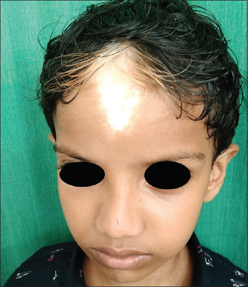
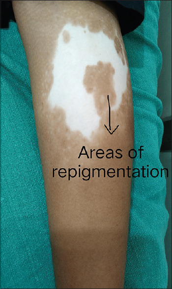
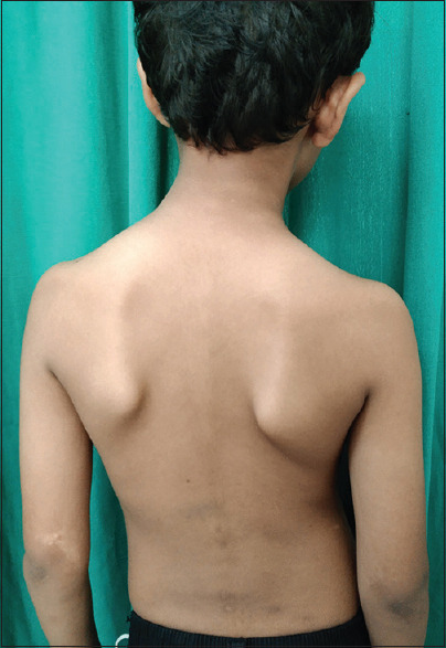
Waardenburg syndrome (WS) is an autosomal dominant genetic disease with an estimated prevalence of 1 in 20,000-40,000. An 8-year-old boy, born to nonconsanguineous parents, presented with complaints of areas of depigmentation on the forehead and right leg since birth. On examination, there was a well-defined amelanotic macule on the center of the forehead of size 5 cm × 2.5 cm with a few spots of normal pigmentation, a forelock of white hair on the frontal area of the scalp, and a well-defined amelanotic macule of size 6 cm × 4 cm with a small central area of normal pigmentation on the posterior part of the right leg. He had scoliosis of the spine in the thoracic region. The nasal root was broad with widely separated inner canthi. There was exotropia and microcornea of the right eye with a visual acuity of 6/24. Fundus examination of the right eye showed a large disc, disc coloboma, peripapillary atrophy, and pigmentary changes in the fovea. The left eye was normal. There was no hearing defect. His father and two siblings too had patchy amelanosis in a similar distribution. They probably represent a limited expression of the same disease. All of them meet the diagnostic criteria for WS. WS is rare with only <100 cases reported worldwide.

 求助内容:
求助内容: 应助结果提醒方式:


