{"title":"瘢痕性脱发的毛病理学。","authors":"Preksha Singh, Taru Garg, Shilpi Agarwal, Amit Kumar Meena, Ram Chander, Deeksha Singh","doi":"10.4103/ijt.ijt_123_23","DOIUrl":null,"url":null,"abstract":"<p><strong>Background: </strong>Patchy/focal alopecia can be noncicatricial or cicatricial. Trichopathology may play an important role in confirming and/or establishing the diagnosis of cicatricial alopecia.</p><p><strong>Aims: </strong>The aim of this study was to study the trichopathologic features of common cicatricial conditions presenting with patchy scalp alopecia.</p><p><strong>Materials and methods: </strong>This cross-sectional study was conducted over a period of 1 year in the department of dermatology and pathology in a tertiary care hospital in North India. Adult patients (age >18 years) presenting with patchy cicatricial alopecia as well as clinically doubtful noncicatricial conditions over the scalp were included in this study. In doubtful cases of noncicatricial alopecia, a single 4-mm punch biopsy was performed for transverse sectioning with hematoxylin and eosin stain. In all cases of cicatricial alopecia, two 4-mm punch biopsies were performed, one for transverse sectioning and the second for vertical sectioning and direct immunofluorescence (DIF). The final diagnosis was made on the basis of the trichopathologic findings.</p><p><strong>Results: </strong>A total of 23 cases were included in our study with the majority (52.2%) being in the age group of 20-30 years. The mean age at diagnosis was 30.5 ± 11.38 years. On the basis of trichopathologic findings, a final diagnosis was made in 18 (78.3%) cases. However, 5 (21.7%) cases remained undiagnosed and were labeled as nonspecific. According to the final diagnosis based on the trichopathologic findings, maximum cases were of discoid lupus erythematosus (DLE) (26.1%), followed by lichen planopilaris (21.7%), pseudopelade of Brocq (13%), alopecia areata (8.7%), folliculitis decalvans (4.3%), and traction alopecia (4.3%). Trichopathologic examination revealed hyperkeratosis (60%), basal cell vacuolization (55%), epidermal atrophy (55%,) perifollicular lymphocytic infiltrate (30%), pigment incontinence (30%), perifollicular fibrosis (25%), and thickened basement membrane (25%). Only four cases showed deposits of immunoreactants in DIF and all four were finally diagnosed as DLE.</p><p><strong>Conclusions: </strong>Trichopathology along with special stain is a useful tool in the diagnosis of patchy alopecia and must be carried out in cases where diagnosis is in doubt clinically, particularly in cases of cicatricial alopecia. DIF may have a supportive role in histopathologically inconclusive cases and should be carried out, particularly when DLE is suspected. Trichopathology in combination with DIF is an important tool in the diagnosis of the majority of cases of cicatricial as well as clinically doubtful cases of noncicatricial alopecia.</p>","PeriodicalId":14417,"journal":{"name":"International Journal of Trichology","volume":"17 1","pages":"58-63"},"PeriodicalIF":0.0000,"publicationDate":"2025-01-01","publicationTypes":"Journal Article","fieldsOfStudy":null,"isOpenAccess":false,"openAccessPdf":"https://www.ncbi.nlm.nih.gov/pmc/articles/PMC12251984/pdf/","citationCount":"0","resultStr":"{\"title\":\"Trichopathology of Cicatricial Alopecia.\",\"authors\":\"Preksha Singh, Taru Garg, Shilpi Agarwal, Amit Kumar Meena, Ram Chander, Deeksha Singh\",\"doi\":\"10.4103/ijt.ijt_123_23\",\"DOIUrl\":null,\"url\":null,\"abstract\":\"<p><strong>Background: </strong>Patchy/focal alopecia can be noncicatricial or cicatricial. Trichopathology may play an important role in confirming and/or establishing the diagnosis of cicatricial alopecia.</p><p><strong>Aims: </strong>The aim of this study was to study the trichopathologic features of common cicatricial conditions presenting with patchy scalp alopecia.</p><p><strong>Materials and methods: </strong>This cross-sectional study was conducted over a period of 1 year in the department of dermatology and pathology in a tertiary care hospital in North India. Adult patients (age >18 years) presenting with patchy cicatricial alopecia as well as clinically doubtful noncicatricial conditions over the scalp were included in this study. In doubtful cases of noncicatricial alopecia, a single 4-mm punch biopsy was performed for transverse sectioning with hematoxylin and eosin stain. In all cases of cicatricial alopecia, two 4-mm punch biopsies were performed, one for transverse sectioning and the second for vertical sectioning and direct immunofluorescence (DIF). The final diagnosis was made on the basis of the trichopathologic findings.</p><p><strong>Results: </strong>A total of 23 cases were included in our study with the majority (52.2%) being in the age group of 20-30 years. The mean age at diagnosis was 30.5 ± 11.38 years. On the basis of trichopathologic findings, a final diagnosis was made in 18 (78.3%) cases. However, 5 (21.7%) cases remained undiagnosed and were labeled as nonspecific. According to the final diagnosis based on the trichopathologic findings, maximum cases were of discoid lupus erythematosus (DLE) (26.1%), followed by lichen planopilaris (21.7%), pseudopelade of Brocq (13%), alopecia areata (8.7%), folliculitis decalvans (4.3%), and traction alopecia (4.3%). Trichopathologic examination revealed hyperkeratosis (60%), basal cell vacuolization (55%), epidermal atrophy (55%,) perifollicular lymphocytic infiltrate (30%), pigment incontinence (30%), perifollicular fibrosis (25%), and thickened basement membrane (25%). Only four cases showed deposits of immunoreactants in DIF and all four were finally diagnosed as DLE.</p><p><strong>Conclusions: </strong>Trichopathology along with special stain is a useful tool in the diagnosis of patchy alopecia and must be carried out in cases where diagnosis is in doubt clinically, particularly in cases of cicatricial alopecia. DIF may have a supportive role in histopathologically inconclusive cases and should be carried out, particularly when DLE is suspected. Trichopathology in combination with DIF is an important tool in the diagnosis of the majority of cases of cicatricial as well as clinically doubtful cases of noncicatricial alopecia.</p>\",\"PeriodicalId\":14417,\"journal\":{\"name\":\"International Journal of Trichology\",\"volume\":\"17 1\",\"pages\":\"58-63\"},\"PeriodicalIF\":0.0000,\"publicationDate\":\"2025-01-01\",\"publicationTypes\":\"Journal Article\",\"fieldsOfStudy\":null,\"isOpenAccess\":false,\"openAccessPdf\":\"https://www.ncbi.nlm.nih.gov/pmc/articles/PMC12251984/pdf/\",\"citationCount\":\"0\",\"resultStr\":null,\"platform\":\"Semanticscholar\",\"paperid\":null,\"PeriodicalName\":\"International Journal of Trichology\",\"FirstCategoryId\":\"1085\",\"ListUrlMain\":\"https://doi.org/10.4103/ijt.ijt_123_23\",\"RegionNum\":0,\"RegionCategory\":null,\"ArticlePicture\":[],\"TitleCN\":null,\"AbstractTextCN\":null,\"PMCID\":null,\"EPubDate\":\"2025/6/23 0:00:00\",\"PubModel\":\"Epub\",\"JCR\":\"Q2\",\"JCRName\":\"Medicine\",\"Score\":null,\"Total\":0}","platform":"Semanticscholar","paperid":null,"PeriodicalName":"International Journal of Trichology","FirstCategoryId":"1085","ListUrlMain":"https://doi.org/10.4103/ijt.ijt_123_23","RegionNum":0,"RegionCategory":null,"ArticlePicture":[],"TitleCN":null,"AbstractTextCN":null,"PMCID":null,"EPubDate":"2025/6/23 0:00:00","PubModel":"Epub","JCR":"Q2","JCRName":"Medicine","Score":null,"Total":0}
Background: Patchy/focal alopecia can be noncicatricial or cicatricial. Trichopathology may play an important role in confirming and/or establishing the diagnosis of cicatricial alopecia.
Aims: The aim of this study was to study the trichopathologic features of common cicatricial conditions presenting with patchy scalp alopecia.
Materials and methods: This cross-sectional study was conducted over a period of 1 year in the department of dermatology and pathology in a tertiary care hospital in North India. Adult patients (age >18 years) presenting with patchy cicatricial alopecia as well as clinically doubtful noncicatricial conditions over the scalp were included in this study. In doubtful cases of noncicatricial alopecia, a single 4-mm punch biopsy was performed for transverse sectioning with hematoxylin and eosin stain. In all cases of cicatricial alopecia, two 4-mm punch biopsies were performed, one for transverse sectioning and the second for vertical sectioning and direct immunofluorescence (DIF). The final diagnosis was made on the basis of the trichopathologic findings.
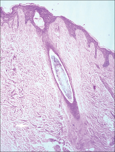
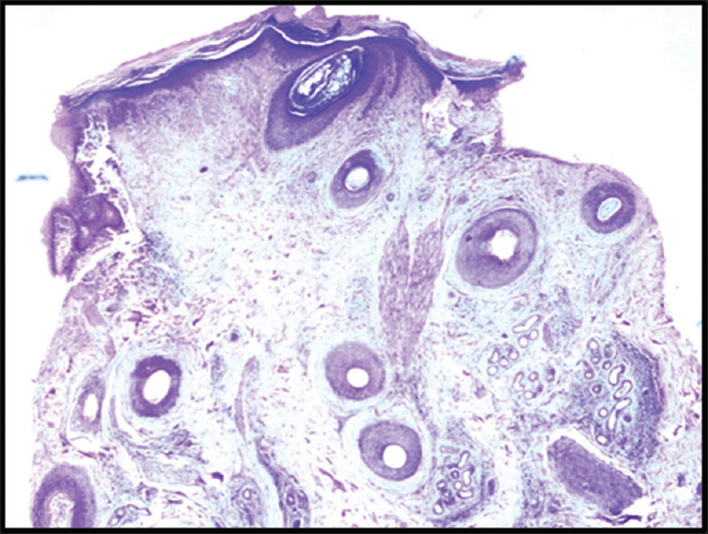
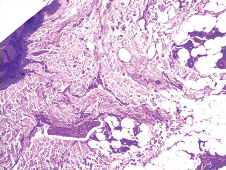
Results: A total of 23 cases were included in our study with the majority (52.2%) being in the age group of 20-30 years. The mean age at diagnosis was 30.5 ± 11.38 years. On the basis of trichopathologic findings, a final diagnosis was made in 18 (78.3%) cases. However, 5 (21.7%) cases remained undiagnosed and were labeled as nonspecific. According to the final diagnosis based on the trichopathologic findings, maximum cases were of discoid lupus erythematosus (DLE) (26.1%), followed by lichen planopilaris (21.7%), pseudopelade of Brocq (13%), alopecia areata (8.7%), folliculitis decalvans (4.3%), and traction alopecia (4.3%). Trichopathologic examination revealed hyperkeratosis (60%), basal cell vacuolization (55%), epidermal atrophy (55%,) perifollicular lymphocytic infiltrate (30%), pigment incontinence (30%), perifollicular fibrosis (25%), and thickened basement membrane (25%). Only four cases showed deposits of immunoreactants in DIF and all four were finally diagnosed as DLE.
Conclusions: Trichopathology along with special stain is a useful tool in the diagnosis of patchy alopecia and must be carried out in cases where diagnosis is in doubt clinically, particularly in cases of cicatricial alopecia. DIF may have a supportive role in histopathologically inconclusive cases and should be carried out, particularly when DLE is suspected. Trichopathology in combination with DIF is an important tool in the diagnosis of the majority of cases of cicatricial as well as clinically doubtful cases of noncicatricial alopecia.

 求助内容:
求助内容: 应助结果提醒方式:


