Feifu Wang, Stephen J Vincent, Pauline Cho, Yi Shen, Zihao Sheng, Meixiao Shen, Jun Jiang
{"title":"短期巩膜晶状体磨损时广角流体储层厚度的变化。","authors":"Feifu Wang, Stephen J Vincent, Pauline Cho, Yi Shen, Zihao Sheng, Meixiao Shen, Jun Jiang","doi":"10.1186/s40662-025-00443-3","DOIUrl":null,"url":null,"abstract":"<p><strong>Background: </strong>To analyze the fluid reservoir thickness over the whole cornea during scleral lens settling using wide-angle optical coherence tomography (OCT) images and customized computer software.</p><p><strong>Methods: </strong>A total of 75 participants were recruited - 29 (myopes) with regular corneas and 46 with irregular corneas (35 with keratoconus, and 11 post-keratoplasty). All participants were fitted with customized scleral lenses and anterior segment OCT (Tomey Casia 2) images were taken 0, 30, 60, 120, and 240 min after lens application at the dispensing visit. Customized software was used to automatically segment the anterior cornea and the posterior surface of the scleral lens and determine the fluid reservoir thickness at 17 corneal regions across a 12 mm diameter.</p><p><strong>Results: </strong>Fluid reservoir thickness decreased over time (P < 0.001) following an exponential decay, with no differences observed over time between the three groups (P = 0.97). The reduction in fluid reservoir thickness over four hours varied slightly between the central (149 ± 9 μm), mid-peripheral (139 ± 11 μm), and peripheral regions (131 ± 15 μm), P = 0.046. The fluid reservoir was thinnest in the superior mid-periphery for both the myopia and post-keratoplasty groups, and centrally for the keratoconus group. The fluid reservoir was thickest inferiorly for all groups, with the greatest level of asymmetry observed along the vertical meridian.</p><p><strong>Conclusions: </strong>Fluid reservoir thickness decreased most rapidly during the first two hours of lens wear and followed an exponential decay for both regular and irregular corneas across all corneal locations. Fluid reservoir asymmetry was greatest along the vertical meridian with a thicker reservoir observed in the inferior corneal regions.</p>","PeriodicalId":12194,"journal":{"name":"Eye and Vision","volume":"12 1","pages":"27"},"PeriodicalIF":4.0000,"publicationDate":"2025-07-14","publicationTypes":"Journal Article","fieldsOfStudy":null,"isOpenAccess":false,"openAccessPdf":"https://www.ncbi.nlm.nih.gov/pmc/articles/PMC12257854/pdf/","citationCount":"0","resultStr":"{\"title\":\"Wide-angle fluid reservoir thickness changes during short-term scleral lens wear.\",\"authors\":\"Feifu Wang, Stephen J Vincent, Pauline Cho, Yi Shen, Zihao Sheng, Meixiao Shen, Jun Jiang\",\"doi\":\"10.1186/s40662-025-00443-3\",\"DOIUrl\":null,\"url\":null,\"abstract\":\"<p><strong>Background: </strong>To analyze the fluid reservoir thickness over the whole cornea during scleral lens settling using wide-angle optical coherence tomography (OCT) images and customized computer software.</p><p><strong>Methods: </strong>A total of 75 participants were recruited - 29 (myopes) with regular corneas and 46 with irregular corneas (35 with keratoconus, and 11 post-keratoplasty). All participants were fitted with customized scleral lenses and anterior segment OCT (Tomey Casia 2) images were taken 0, 30, 60, 120, and 240 min after lens application at the dispensing visit. Customized software was used to automatically segment the anterior cornea and the posterior surface of the scleral lens and determine the fluid reservoir thickness at 17 corneal regions across a 12 mm diameter.</p><p><strong>Results: </strong>Fluid reservoir thickness decreased over time (P < 0.001) following an exponential decay, with no differences observed over time between the three groups (P = 0.97). The reduction in fluid reservoir thickness over four hours varied slightly between the central (149 ± 9 μm), mid-peripheral (139 ± 11 μm), and peripheral regions (131 ± 15 μm), P = 0.046. The fluid reservoir was thinnest in the superior mid-periphery for both the myopia and post-keratoplasty groups, and centrally for the keratoconus group. The fluid reservoir was thickest inferiorly for all groups, with the greatest level of asymmetry observed along the vertical meridian.</p><p><strong>Conclusions: </strong>Fluid reservoir thickness decreased most rapidly during the first two hours of lens wear and followed an exponential decay for both regular and irregular corneas across all corneal locations. Fluid reservoir asymmetry was greatest along the vertical meridian with a thicker reservoir observed in the inferior corneal regions.</p>\",\"PeriodicalId\":12194,\"journal\":{\"name\":\"Eye and Vision\",\"volume\":\"12 1\",\"pages\":\"27\"},\"PeriodicalIF\":4.0000,\"publicationDate\":\"2025-07-14\",\"publicationTypes\":\"Journal Article\",\"fieldsOfStudy\":null,\"isOpenAccess\":false,\"openAccessPdf\":\"https://www.ncbi.nlm.nih.gov/pmc/articles/PMC12257854/pdf/\",\"citationCount\":\"0\",\"resultStr\":null,\"platform\":\"Semanticscholar\",\"paperid\":null,\"PeriodicalName\":\"Eye and Vision\",\"FirstCategoryId\":\"3\",\"ListUrlMain\":\"https://doi.org/10.1186/s40662-025-00443-3\",\"RegionNum\":1,\"RegionCategory\":\"医学\",\"ArticlePicture\":[],\"TitleCN\":null,\"AbstractTextCN\":null,\"PMCID\":null,\"EPubDate\":\"\",\"PubModel\":\"\",\"JCR\":\"Q1\",\"JCRName\":\"OPHTHALMOLOGY\",\"Score\":null,\"Total\":0}","platform":"Semanticscholar","paperid":null,"PeriodicalName":"Eye and Vision","FirstCategoryId":"3","ListUrlMain":"https://doi.org/10.1186/s40662-025-00443-3","RegionNum":1,"RegionCategory":"医学","ArticlePicture":[],"TitleCN":null,"AbstractTextCN":null,"PMCID":null,"EPubDate":"","PubModel":"","JCR":"Q1","JCRName":"OPHTHALMOLOGY","Score":null,"Total":0}
Wide-angle fluid reservoir thickness changes during short-term scleral lens wear.
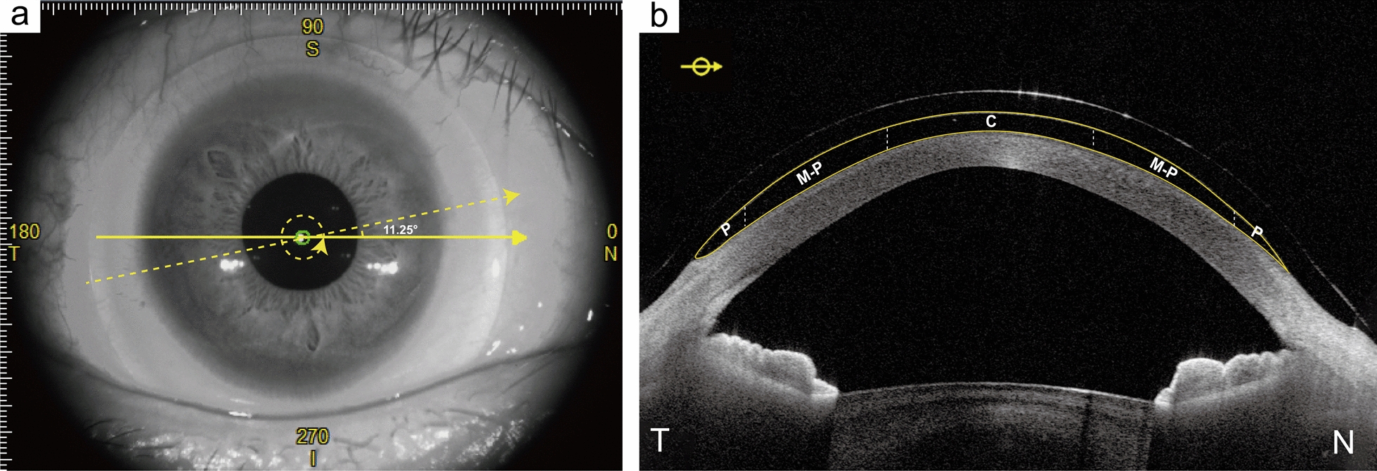
Background: To analyze the fluid reservoir thickness over the whole cornea during scleral lens settling using wide-angle optical coherence tomography (OCT) images and customized computer software.
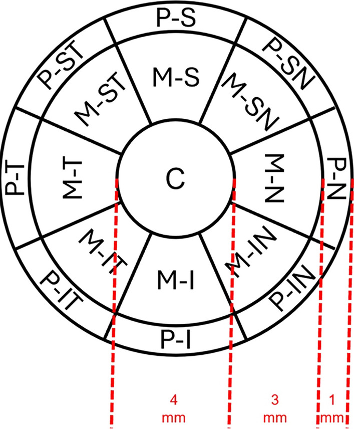
Methods: A total of 75 participants were recruited - 29 (myopes) with regular corneas and 46 with irregular corneas (35 with keratoconus, and 11 post-keratoplasty). All participants were fitted with customized scleral lenses and anterior segment OCT (Tomey Casia 2) images were taken 0, 30, 60, 120, and 240 min after lens application at the dispensing visit. Customized software was used to automatically segment the anterior cornea and the posterior surface of the scleral lens and determine the fluid reservoir thickness at 17 corneal regions across a 12 mm diameter.
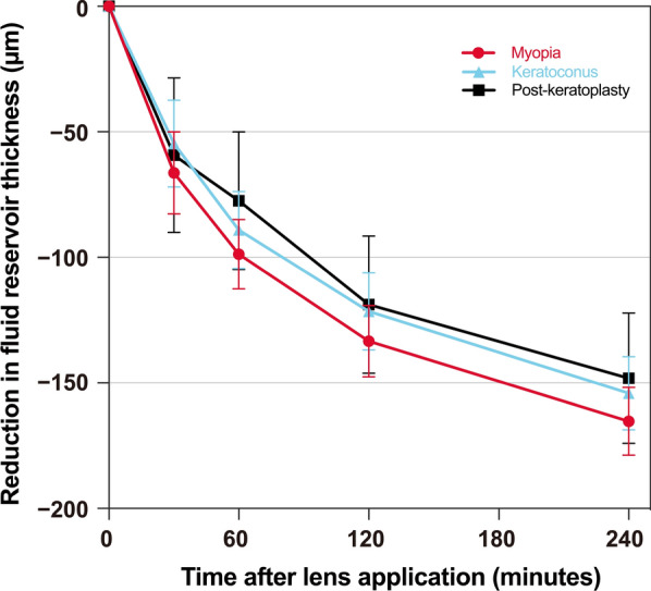
Results: Fluid reservoir thickness decreased over time (P < 0.001) following an exponential decay, with no differences observed over time between the three groups (P = 0.97). The reduction in fluid reservoir thickness over four hours varied slightly between the central (149 ± 9 μm), mid-peripheral (139 ± 11 μm), and peripheral regions (131 ± 15 μm), P = 0.046. The fluid reservoir was thinnest in the superior mid-periphery for both the myopia and post-keratoplasty groups, and centrally for the keratoconus group. The fluid reservoir was thickest inferiorly for all groups, with the greatest level of asymmetry observed along the vertical meridian.
Conclusions: Fluid reservoir thickness decreased most rapidly during the first two hours of lens wear and followed an exponential decay for both regular and irregular corneas across all corneal locations. Fluid reservoir asymmetry was greatest along the vertical meridian with a thicker reservoir observed in the inferior corneal regions.
期刊介绍:
Eye and Vision is an open access, peer-reviewed journal for ophthalmologists and visual science specialists. It welcomes research articles, reviews, methodologies, commentaries, case reports, perspectives and short reports encompassing all aspects of eye and vision. Topics of interest include but are not limited to: current developments of theoretical, experimental and clinical investigations in ophthalmology, optometry and vision science which focus on novel and high-impact findings on central issues pertaining to biology, pathophysiology and etiology of eye diseases as well as advances in diagnostic techniques, surgical treatment, instrument updates, the latest drug findings, results of clinical trials and research findings. It aims to provide ophthalmologists and visual science specialists with the latest developments in theoretical, experimental and clinical investigations in eye and vision.
 求助内容:
求助内容: 应助结果提醒方式:


