Jiawen Xu, Yihua Jin, Min Wang, Yijing Tao, Yujia Wang, Fangqi Gong
{"title":"双调节蛋白促进川崎病细胞模型中受损内皮细胞的增殖和迁移","authors":"Jiawen Xu, Yihua Jin, Min Wang, Yijing Tao, Yujia Wang, Fangqi Gong","doi":"10.1002/iid3.70223","DOIUrl":null,"url":null,"abstract":"<div>\n \n \n <section>\n \n <h3> Objectives</h3>\n \n <p>Amphiregulin (Areg), a member of the epidermal growth factor family, plays a critical role in tissue repair, inflammation, and immunity. Macrophages are an important source of Areg and are also among the key immune cells activated in Kawasaki disease (KD). Despite this, the role of Areg in KD has not been studied. Therefore, this study aims to investigate the expression of Areg in a KD model and to elucidate its effects on injured endothelial cells using a KD cell model.</p>\n </section>\n \n <section>\n \n <h3> Methods</h3>\n \n <p>The serum of LCWE-induced KD mouse model was measured by ELISA. RAW264.7 cells were stimulated with LCWE, and the supernatant was collected. Then, MCAECs were treated with LCWE-induced RAW264.7 cells conditioned medium (RAW-CM) to simulate inflammatory damage in KD endothelial cells.</p>\n </section>\n \n <section>\n \n <h3> Results</h3>\n \n <p>Our study showed that the serum level of Areg increased in LCWE-induced mouse model. In vitro, LCWE increased the expression and secretion of Areg in RAW264.7 macrophages, an effect that was inhibited by ADAM-17 blockade. The conditioned medium (CM) from LCWE-stimulated RAW264.7 cells (RAW-CM) enhanced the proliferative capacity of endothelial cells, an effect that was partially inhibited by Areg antibodies. Recombinant Areg promoted the proliferation and migration of damaged endothelial cells, effects that were dependent on the activation of the AKT and ERK signaling pathways.</p>\n </section>\n \n <section>\n \n <h3> Conclusion</h3>\n \n <p>This study demonstrates that serum Areg level increased in LCWE-induced KD mouse model, and Areg promoted proliferation and migration abilities of injured endothelial cells. Our work suggests that Areg may be one of the reasons for the repair of injured endothelial cells in LCWE model vasculitis.</p>\n </section>\n </div>","PeriodicalId":13289,"journal":{"name":"Immunity, Inflammation and Disease","volume":"13 7","pages":""},"PeriodicalIF":2.7000,"publicationDate":"2025-07-15","publicationTypes":"Journal Article","fieldsOfStudy":null,"isOpenAccess":false,"openAccessPdf":"https://onlinelibrary.wiley.com/doi/epdf/10.1002/iid3.70223","citationCount":"0","resultStr":"{\"title\":\"Amphiregulin Promotes Proliferation and Migration of the Damaged Endothelial Cells in Kawasaki Disease Cell Models\",\"authors\":\"Jiawen Xu, Yihua Jin, Min Wang, Yijing Tao, Yujia Wang, Fangqi Gong\",\"doi\":\"10.1002/iid3.70223\",\"DOIUrl\":null,\"url\":null,\"abstract\":\"<div>\\n \\n \\n <section>\\n \\n <h3> Objectives</h3>\\n \\n <p>Amphiregulin (Areg), a member of the epidermal growth factor family, plays a critical role in tissue repair, inflammation, and immunity. Macrophages are an important source of Areg and are also among the key immune cells activated in Kawasaki disease (KD). Despite this, the role of Areg in KD has not been studied. Therefore, this study aims to investigate the expression of Areg in a KD model and to elucidate its effects on injured endothelial cells using a KD cell model.</p>\\n </section>\\n \\n <section>\\n \\n <h3> Methods</h3>\\n \\n <p>The serum of LCWE-induced KD mouse model was measured by ELISA. RAW264.7 cells were stimulated with LCWE, and the supernatant was collected. Then, MCAECs were treated with LCWE-induced RAW264.7 cells conditioned medium (RAW-CM) to simulate inflammatory damage in KD endothelial cells.</p>\\n </section>\\n \\n <section>\\n \\n <h3> Results</h3>\\n \\n <p>Our study showed that the serum level of Areg increased in LCWE-induced mouse model. In vitro, LCWE increased the expression and secretion of Areg in RAW264.7 macrophages, an effect that was inhibited by ADAM-17 blockade. The conditioned medium (CM) from LCWE-stimulated RAW264.7 cells (RAW-CM) enhanced the proliferative capacity of endothelial cells, an effect that was partially inhibited by Areg antibodies. Recombinant Areg promoted the proliferation and migration of damaged endothelial cells, effects that were dependent on the activation of the AKT and ERK signaling pathways.</p>\\n </section>\\n \\n <section>\\n \\n <h3> Conclusion</h3>\\n \\n <p>This study demonstrates that serum Areg level increased in LCWE-induced KD mouse model, and Areg promoted proliferation and migration abilities of injured endothelial cells. Our work suggests that Areg may be one of the reasons for the repair of injured endothelial cells in LCWE model vasculitis.</p>\\n </section>\\n </div>\",\"PeriodicalId\":13289,\"journal\":{\"name\":\"Immunity, Inflammation and Disease\",\"volume\":\"13 7\",\"pages\":\"\"},\"PeriodicalIF\":2.7000,\"publicationDate\":\"2025-07-15\",\"publicationTypes\":\"Journal Article\",\"fieldsOfStudy\":null,\"isOpenAccess\":false,\"openAccessPdf\":\"https://onlinelibrary.wiley.com/doi/epdf/10.1002/iid3.70223\",\"citationCount\":\"0\",\"resultStr\":null,\"platform\":\"Semanticscholar\",\"paperid\":null,\"PeriodicalName\":\"Immunity, Inflammation and Disease\",\"FirstCategoryId\":\"3\",\"ListUrlMain\":\"https://onlinelibrary.wiley.com/doi/10.1002/iid3.70223\",\"RegionNum\":4,\"RegionCategory\":\"医学\",\"ArticlePicture\":[],\"TitleCN\":null,\"AbstractTextCN\":null,\"PMCID\":null,\"EPubDate\":\"\",\"PubModel\":\"\",\"JCR\":\"Q3\",\"JCRName\":\"IMMUNOLOGY\",\"Score\":null,\"Total\":0}","platform":"Semanticscholar","paperid":null,"PeriodicalName":"Immunity, Inflammation and Disease","FirstCategoryId":"3","ListUrlMain":"https://onlinelibrary.wiley.com/doi/10.1002/iid3.70223","RegionNum":4,"RegionCategory":"医学","ArticlePicture":[],"TitleCN":null,"AbstractTextCN":null,"PMCID":null,"EPubDate":"","PubModel":"","JCR":"Q3","JCRName":"IMMUNOLOGY","Score":null,"Total":0}
Amphiregulin Promotes Proliferation and Migration of the Damaged Endothelial Cells in Kawasaki Disease Cell Models
Objectives
Amphiregulin (Areg), a member of the epidermal growth factor family, plays a critical role in tissue repair, inflammation, and immunity. Macrophages are an important source of Areg and are also among the key immune cells activated in Kawasaki disease (KD). Despite this, the role of Areg in KD has not been studied. Therefore, this study aims to investigate the expression of Areg in a KD model and to elucidate its effects on injured endothelial cells using a KD cell model.
Methods
The serum of LCWE-induced KD mouse model was measured by ELISA. RAW264.7 cells were stimulated with LCWE, and the supernatant was collected. Then, MCAECs were treated with LCWE-induced RAW264.7 cells conditioned medium (RAW-CM) to simulate inflammatory damage in KD endothelial cells.
Results
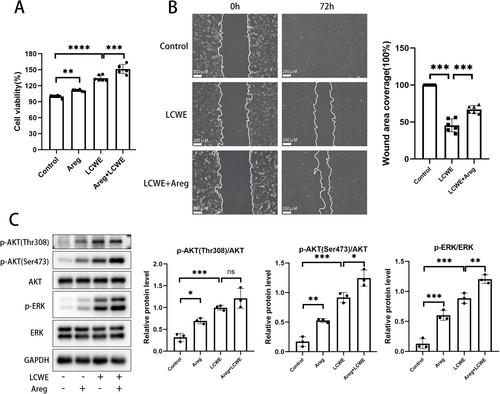
Our study showed that the serum level of Areg increased in LCWE-induced mouse model. In vitro, LCWE increased the expression and secretion of Areg in RAW264.7 macrophages, an effect that was inhibited by ADAM-17 blockade. The conditioned medium (CM) from LCWE-stimulated RAW264.7 cells (RAW-CM) enhanced the proliferative capacity of endothelial cells, an effect that was partially inhibited by Areg antibodies. Recombinant Areg promoted the proliferation and migration of damaged endothelial cells, effects that were dependent on the activation of the AKT and ERK signaling pathways.
Conclusion
This study demonstrates that serum Areg level increased in LCWE-induced KD mouse model, and Areg promoted proliferation and migration abilities of injured endothelial cells. Our work suggests that Areg may be one of the reasons for the repair of injured endothelial cells in LCWE model vasculitis.
期刊介绍:
Immunity, Inflammation and Disease is a peer-reviewed, open access, interdisciplinary journal providing rapid publication of research across the broad field of immunology. Immunity, Inflammation and Disease gives rapid consideration to papers in all areas of clinical and basic research. The journal is indexed in Medline and the Science Citation Index Expanded (part of Web of Science), among others. It welcomes original work that enhances the understanding of immunology in areas including:
• cellular and molecular immunology
• clinical immunology
• allergy
• immunochemistry
• immunogenetics
• immune signalling
• immune development
• imaging
• mathematical modelling
• autoimmunity
• transplantation immunology
• cancer immunology
 求助内容:
求助内容: 应助结果提醒方式:


