Sahil Shet, Catherine Henry, Mairead O’Donnell, Eid Kakish, Muhammad Ghauri, Patrick O’Regan, Kevin Deasy, Hisham Ibrahim, Michael Maher, Barry Plant, David J. Ryan
{"title":"利用超低剂量胸部CT评估囊性纤维化患者的骨密度。","authors":"Sahil Shet, Catherine Henry, Mairead O’Donnell, Eid Kakish, Muhammad Ghauri, Patrick O’Regan, Kevin Deasy, Hisham Ibrahim, Michael Maher, Barry Plant, David J. Ryan","doi":"10.1007/s11657-025-01578-5","DOIUrl":null,"url":null,"abstract":"<p>In this study, we used routine ultra-low dose computed tomography scans of patients with cystic fibrosis to predict bone mineral density (BMD). A strong correlation was found between the attenuation of trabecular bone in thoracic vertebrae and the BMD in the proximal femur and lumbar spine as measured on DEXA.</p><p>Osteoporosis is a serious global health concern with millions of people affected worldwide. A particularly vulnerable cohort in developing osteoporosis are patients with cystic fibrosis (CF). Bone mineral density (BMD) is typically measured with dual energy X-ray absorptiometry (DEXA) scanning; however, this comes at a cost to the healthcare system and an exposure to ionising radiation. In our institution, patients with cystic fibrosis undergo routine ultra-low dose computed tomography (ULDCT) for monitoring of disease progression. The aim of this study was to assess the validity of estimating BMD using data derived from ULDCT scans.</p><p>Adult CF patients were included if they had undergone a routine ULDCT scan within 12 months of a DEXA scan. Additionally, 100 non-CF patients with non-contrast standard dose CT scans were selected to act as the control group. Trabecular bone density (T-BD) at T4, T7 and T10 was measured on PACS in Hounsfield units (HU) and compared to DEXA scan results and a formula developed to the predict BMD.</p><p>Fifty-two female and 62 male patients were included with mean ages of 34.4 and 35.1 respectively. Moderately strong correlation was found between the T-BD and BMD of both the lumbar spine (<i>r</i> = 0.629, <i>p</i> < 0.001) and proximal femur (<i>r</i> = 0.649, <i>p</i> < 0.001). Receiver operator characteristic (ROC) curve analysis found a sensitivity and specificity of 0.700 and 0.714 respectively at predicting osteoporosis at T-BD of 193.33 HU or below.</p><p>T-BD measured on ULDCT may be a valuable tool in the early identification of CF patients at risk of osteoporosis.\n</p>","PeriodicalId":8283,"journal":{"name":"Archives of Osteoporosis","volume":"20 1","pages":""},"PeriodicalIF":2.8000,"publicationDate":"2025-07-12","publicationTypes":"Journal Article","fieldsOfStudy":null,"isOpenAccess":false,"openAccessPdf":"https://www.ncbi.nlm.nih.gov/pmc/articles/PMC12255546/pdf/","citationCount":"0","resultStr":"{\"title\":\"Opportunistic assessment of bone mineral density in cystic fibrosis patients using ultra-low dose thoracic CT\",\"authors\":\"Sahil Shet, Catherine Henry, Mairead O’Donnell, Eid Kakish, Muhammad Ghauri, Patrick O’Regan, Kevin Deasy, Hisham Ibrahim, Michael Maher, Barry Plant, David J. Ryan\",\"doi\":\"10.1007/s11657-025-01578-5\",\"DOIUrl\":null,\"url\":null,\"abstract\":\"<p>In this study, we used routine ultra-low dose computed tomography scans of patients with cystic fibrosis to predict bone mineral density (BMD). A strong correlation was found between the attenuation of trabecular bone in thoracic vertebrae and the BMD in the proximal femur and lumbar spine as measured on DEXA.</p><p>Osteoporosis is a serious global health concern with millions of people affected worldwide. A particularly vulnerable cohort in developing osteoporosis are patients with cystic fibrosis (CF). Bone mineral density (BMD) is typically measured with dual energy X-ray absorptiometry (DEXA) scanning; however, this comes at a cost to the healthcare system and an exposure to ionising radiation. In our institution, patients with cystic fibrosis undergo routine ultra-low dose computed tomography (ULDCT) for monitoring of disease progression. The aim of this study was to assess the validity of estimating BMD using data derived from ULDCT scans.</p><p>Adult CF patients were included if they had undergone a routine ULDCT scan within 12 months of a DEXA scan. Additionally, 100 non-CF patients with non-contrast standard dose CT scans were selected to act as the control group. Trabecular bone density (T-BD) at T4, T7 and T10 was measured on PACS in Hounsfield units (HU) and compared to DEXA scan results and a formula developed to the predict BMD.</p><p>Fifty-two female and 62 male patients were included with mean ages of 34.4 and 35.1 respectively. Moderately strong correlation was found between the T-BD and BMD of both the lumbar spine (<i>r</i> = 0.629, <i>p</i> < 0.001) and proximal femur (<i>r</i> = 0.649, <i>p</i> < 0.001). Receiver operator characteristic (ROC) curve analysis found a sensitivity and specificity of 0.700 and 0.714 respectively at predicting osteoporosis at T-BD of 193.33 HU or below.</p><p>T-BD measured on ULDCT may be a valuable tool in the early identification of CF patients at risk of osteoporosis.\\n</p>\",\"PeriodicalId\":8283,\"journal\":{\"name\":\"Archives of Osteoporosis\",\"volume\":\"20 1\",\"pages\":\"\"},\"PeriodicalIF\":2.8000,\"publicationDate\":\"2025-07-12\",\"publicationTypes\":\"Journal Article\",\"fieldsOfStudy\":null,\"isOpenAccess\":false,\"openAccessPdf\":\"https://www.ncbi.nlm.nih.gov/pmc/articles/PMC12255546/pdf/\",\"citationCount\":\"0\",\"resultStr\":null,\"platform\":\"Semanticscholar\",\"paperid\":null,\"PeriodicalName\":\"Archives of Osteoporosis\",\"FirstCategoryId\":\"3\",\"ListUrlMain\":\"https://link.springer.com/article/10.1007/s11657-025-01578-5\",\"RegionNum\":3,\"RegionCategory\":\"医学\",\"ArticlePicture\":[],\"TitleCN\":null,\"AbstractTextCN\":null,\"PMCID\":null,\"EPubDate\":\"\",\"PubModel\":\"\",\"JCR\":\"Q2\",\"JCRName\":\"ENDOCRINOLOGY & METABOLISM\",\"Score\":null,\"Total\":0}","platform":"Semanticscholar","paperid":null,"PeriodicalName":"Archives of Osteoporosis","FirstCategoryId":"3","ListUrlMain":"https://link.springer.com/article/10.1007/s11657-025-01578-5","RegionNum":3,"RegionCategory":"医学","ArticlePicture":[],"TitleCN":null,"AbstractTextCN":null,"PMCID":null,"EPubDate":"","PubModel":"","JCR":"Q2","JCRName":"ENDOCRINOLOGY & METABOLISM","Score":null,"Total":0}
Opportunistic assessment of bone mineral density in cystic fibrosis patients using ultra-low dose thoracic CT
In this study, we used routine ultra-low dose computed tomography scans of patients with cystic fibrosis to predict bone mineral density (BMD). A strong correlation was found between the attenuation of trabecular bone in thoracic vertebrae and the BMD in the proximal femur and lumbar spine as measured on DEXA.
Osteoporosis is a serious global health concern with millions of people affected worldwide. A particularly vulnerable cohort in developing osteoporosis are patients with cystic fibrosis (CF). Bone mineral density (BMD) is typically measured with dual energy X-ray absorptiometry (DEXA) scanning; however, this comes at a cost to the healthcare system and an exposure to ionising radiation. In our institution, patients with cystic fibrosis undergo routine ultra-low dose computed tomography (ULDCT) for monitoring of disease progression. The aim of this study was to assess the validity of estimating BMD using data derived from ULDCT scans.
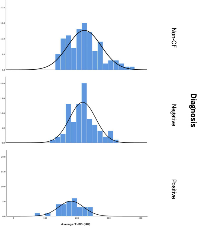
Adult CF patients were included if they had undergone a routine ULDCT scan within 12 months of a DEXA scan. Additionally, 100 non-CF patients with non-contrast standard dose CT scans were selected to act as the control group. Trabecular bone density (T-BD) at T4, T7 and T10 was measured on PACS in Hounsfield units (HU) and compared to DEXA scan results and a formula developed to the predict BMD.
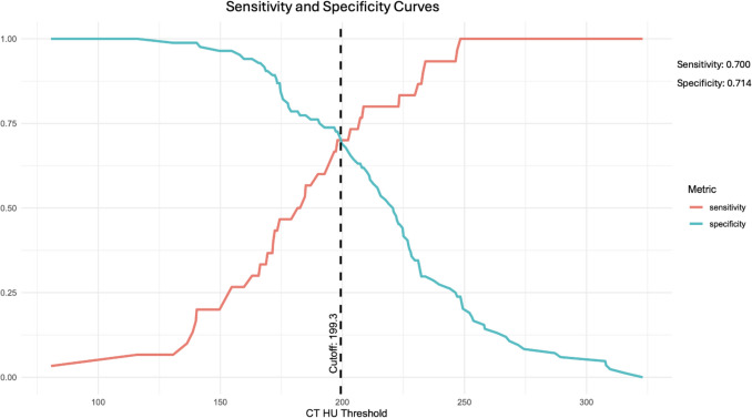
Fifty-two female and 62 male patients were included with mean ages of 34.4 and 35.1 respectively. Moderately strong correlation was found between the T-BD and BMD of both the lumbar spine (r = 0.629, p < 0.001) and proximal femur (r = 0.649, p < 0.001). Receiver operator characteristic (ROC) curve analysis found a sensitivity and specificity of 0.700 and 0.714 respectively at predicting osteoporosis at T-BD of 193.33 HU or below.
T-BD measured on ULDCT may be a valuable tool in the early identification of CF patients at risk of osteoporosis.
期刊介绍:
Archives of Osteoporosis is an international multidisciplinary journal which is a joint initiative of the International Osteoporosis Foundation and the National Osteoporosis Foundation of the USA. The journal will highlight the specificities of different regions around the world concerning epidemiology, reference values for bone density and bone metabolism, as well as clinical aspects of osteoporosis and other bone diseases.
 求助内容:
求助内容: 应助结果提醒方式:


