多模态质谱-拉曼成像揭示溶剂依赖性结构和化学组织改变
IF 6
2区 化学
Q1 CHEMISTRY, ANALYTICAL
引用次数: 0
摘要
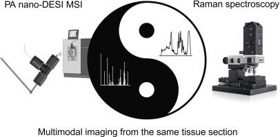
质谱成像(MSI)和拉曼光谱成像是两种化学成像技术,可以显示和可视化薄组织切片中的化学分布。这两种技术是互补的,拉曼光谱检测分子键的拉曼位移,而MSI检测电离分子的质量电荷比。此外,MSI要求分子从表面被解吸、烧蚀或溅射,相比之下使拉曼光谱的破坏性更小。通过多模态分析工作流程,两种技术的优势可以结合起来产生增强的表面化学信息,如薄组织切片。在这里,我们将两种成像方式结合起来,并在薄组织切片的完全相同位置绘制化学特征,以揭示组织中MSI衍生的化学变化。结果在本研究中,我们提出了在同一组织切片上集成气动辅助纳米喷雾解吸电喷雾电离(PA - nano-DESI) MSI和拉曼光谱的多模式工作流程。这就要求MSI对拉曼的采样限制进行一定的调整。我们展示了成功的整合和联合分析在组织化学表征中的力量。事实证明,拉曼光谱对鉴定甲醇或乙腈基溶剂引起的PA纳米desi MSI在组织中引起的溶剂性化学变化非常有效。首先,我们描述了与不同采样模式相关的化学变化,例如不同的探针速度和过采样。接下来,我们描述了暴露于多达五次重复成像实验的所得组织的化学变化。两种方法的结果非常一致,并且表明甲醇比乙腈从组织中脱吸更多的物质,并且两种溶剂都会导致组织中的蛋白质变性。本研究提出了一个多模式的工作流程,结合成像与PA纳米desi MSI和拉曼光谱从完全相同的组织切片和位置。该组合使MSI后组织的化学表征成为可能,并揭示了以前未知的MSI后组织化学的变化。总的来说,我们的研究结果为暴露于溶剂如何影响组织化学提供了新的见解,并强调了将MSI与拉曼光谱相结合用于组织切片深度化学表征的潜力。本文章由计算机程序翻译,如有差异,请以英文原文为准。

Multimodal mass spectrometry - Raman imaging reveals solvent dependent structural and chemical tissue alterations
Background
Mass spectrometry imaging (MSI) and Raman spectroscopy imaging are two chemical imaging techniques that can reveal and visualize the chemical distributions in thin tissue sections. The two techniques are complementary with Raman spectroscopy detecting the Raman shift of molecular bonds while MSI detects mass-to-charge of ionized molecules. Furthermore, MSI requires molecules to be desorbed, ablated or sputtered from the surface, making Raman spectroscopy less destructive in comparison. With a multimodal analysis workflow, the advantages of both techniques can be combined to yield enhanced chemical information of surfaces, such as thin tissue sections. Here, we combine the two imaging modalities and map the chemical signature in the exact same location on thin tissue section to uncover MSI derived chemical alterations in tissue.
Results
In this study, we present a multimodal workflow integrating pneumatically assisted nanospray desorption electrospray ionization (PA nano-DESI) MSI and Raman spectroscopy on the same tissue section. This requires some adaptation of MSI to the sample restrictions of Raman. We demonstrate successful integration and the power of combined analysis in characterization of tissue chemistry. Raman spectroscopy proved highly effective for identifying solvent-induced chemical alterations in tissue caused by PA nano-DESI MSI with methanol or acetonitrile-based solvents. First, we describe chemical alterations connected to different sampling modes, such as different probe speeds and oversampling. Following, we characterize chemical changes in the resulting tissue exposed to up to five repeated imaging experiments. The results from the two modalities were in excellent agreement and showed that methanol desorbs more material from the tissue compared to acetonitrile, and that both solvents cause protein denaturation in the tissue.
Significance
This study presents the establishment of a multimodal workflow combining imaging with PA nano-DESI MSI and Raman spectroscopy from the exact same tissue sections and locations. The combination enabled chemical characterization of the tissue after MSI and revealed previously unknown alterations in tissue chemistry upon MSI. Overall, our findings offer new insights into how exposure to solvents impact tissue chemistry and highlight the potential of combining MSI with Raman spectroscopy for in depth chemical characterization of tissue sections.
求助全文
通过发布文献求助,成功后即可免费获取论文全文。
去求助
来源期刊
Analytica Chimica Acta
化学-分析化学
CiteScore
10.40
自引率
6.50%
发文量
1081
审稿时长
38 days
期刊介绍:
Analytica Chimica Acta has an open access mirror journal Analytica Chimica Acta: X, sharing the same aims and scope, editorial team, submission system and rigorous peer review.
Analytica Chimica Acta provides a forum for the rapid publication of original research, and critical, comprehensive reviews dealing with all aspects of fundamental and applied modern analytical chemistry. The journal welcomes the submission of research papers which report studies concerning the development of new and significant analytical methodologies. In determining the suitability of submitted articles for publication, particular scrutiny will be placed on the degree of novelty and impact of the research and the extent to which it adds to the existing body of knowledge in analytical chemistry.
 求助内容:
求助内容: 应助结果提醒方式:


