Andrzej Fedak, Agnieszka Czapska, Jan Jamroś, Monika Stępień, Tadeusz Popiela
{"title":"超声造影在肝活检中的应用。","authors":"Andrzej Fedak, Agnieszka Czapska, Jan Jamroś, Monika Stępień, Tadeusz Popiela","doi":"10.5114/pjr/203333","DOIUrl":null,"url":null,"abstract":"<p><p>The aim of this study is to determine the usefulness of contrast-enhanced ultrasound (CEUS) in liver biopsy. The popularisation of imaging techniques that visualise the abdominal cavity, especially ultrasonography (USG), has resulted in an increase in the detection of focal liver lesions (FLL). If the results of other imaging modalities (magnetic resonance imaging [MRI] or computed tomography [CT]) are inconclusive, percutaneous liver biopsy should be considered. Taking into account the limitations of using MRI and CT in liver biopsy, this procedure is mostly performed with ultrasound. It is economical, safe, and swift. Whenever it is impossible to visualise lesions in B-mode (a condition necessary for a safe and effective biopsy), it is advisable to use advanced ultrasound techniques - CEUS or fusion imaging. Limitations of fusion imaging include prolonged time of data processing and difficulties in achieving optimal overlap of images. Conversely, CEUS enhances lesion visualisation but is devoid of the mentioned limitations - it is rapid and requires no additional processing. Furthermore, considering the potential of CEUS in the visualisation of focal liver lesions and differentiation of necrotic areas, accompanied by the ability to detect neuroendocrine tumours or its metastasis, we strongly believe that biopsy procedures - especially core needle biopsies - with CEUS assistance are potent tools in contemporary diagnostics. In this paper we want to share the experience of our centre and review the available literature on performing liver biopsies under CEUS guidance.</p>","PeriodicalId":94174,"journal":{"name":"Polish journal of radiology","volume":"90 ","pages":"e292-e298"},"PeriodicalIF":0.0000,"publicationDate":"2025-06-06","publicationTypes":"Journal Article","fieldsOfStudy":null,"isOpenAccess":false,"openAccessPdf":"https://www.ncbi.nlm.nih.gov/pmc/articles/PMC12243522/pdf/","citationCount":"0","resultStr":"{\"title\":\"Application of contrast-enhanced ultrasound in liver biopsy.\",\"authors\":\"Andrzej Fedak, Agnieszka Czapska, Jan Jamroś, Monika Stępień, Tadeusz Popiela\",\"doi\":\"10.5114/pjr/203333\",\"DOIUrl\":null,\"url\":null,\"abstract\":\"<p><p>The aim of this study is to determine the usefulness of contrast-enhanced ultrasound (CEUS) in liver biopsy. The popularisation of imaging techniques that visualise the abdominal cavity, especially ultrasonography (USG), has resulted in an increase in the detection of focal liver lesions (FLL). If the results of other imaging modalities (magnetic resonance imaging [MRI] or computed tomography [CT]) are inconclusive, percutaneous liver biopsy should be considered. Taking into account the limitations of using MRI and CT in liver biopsy, this procedure is mostly performed with ultrasound. It is economical, safe, and swift. Whenever it is impossible to visualise lesions in B-mode (a condition necessary for a safe and effective biopsy), it is advisable to use advanced ultrasound techniques - CEUS or fusion imaging. Limitations of fusion imaging include prolonged time of data processing and difficulties in achieving optimal overlap of images. Conversely, CEUS enhances lesion visualisation but is devoid of the mentioned limitations - it is rapid and requires no additional processing. Furthermore, considering the potential of CEUS in the visualisation of focal liver lesions and differentiation of necrotic areas, accompanied by the ability to detect neuroendocrine tumours or its metastasis, we strongly believe that biopsy procedures - especially core needle biopsies - with CEUS assistance are potent tools in contemporary diagnostics. In this paper we want to share the experience of our centre and review the available literature on performing liver biopsies under CEUS guidance.</p>\",\"PeriodicalId\":94174,\"journal\":{\"name\":\"Polish journal of radiology\",\"volume\":\"90 \",\"pages\":\"e292-e298\"},\"PeriodicalIF\":0.0000,\"publicationDate\":\"2025-06-06\",\"publicationTypes\":\"Journal Article\",\"fieldsOfStudy\":null,\"isOpenAccess\":false,\"openAccessPdf\":\"https://www.ncbi.nlm.nih.gov/pmc/articles/PMC12243522/pdf/\",\"citationCount\":\"0\",\"resultStr\":null,\"platform\":\"Semanticscholar\",\"paperid\":null,\"PeriodicalName\":\"Polish journal of radiology\",\"FirstCategoryId\":\"1085\",\"ListUrlMain\":\"https://doi.org/10.5114/pjr/203333\",\"RegionNum\":0,\"RegionCategory\":null,\"ArticlePicture\":[],\"TitleCN\":null,\"AbstractTextCN\":null,\"PMCID\":null,\"EPubDate\":\"2025/1/1 0:00:00\",\"PubModel\":\"eCollection\",\"JCR\":\"\",\"JCRName\":\"\",\"Score\":null,\"Total\":0}","platform":"Semanticscholar","paperid":null,"PeriodicalName":"Polish journal of radiology","FirstCategoryId":"1085","ListUrlMain":"https://doi.org/10.5114/pjr/203333","RegionNum":0,"RegionCategory":null,"ArticlePicture":[],"TitleCN":null,"AbstractTextCN":null,"PMCID":null,"EPubDate":"2025/1/1 0:00:00","PubModel":"eCollection","JCR":"","JCRName":"","Score":null,"Total":0}
Application of contrast-enhanced ultrasound in liver biopsy.
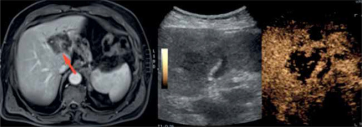
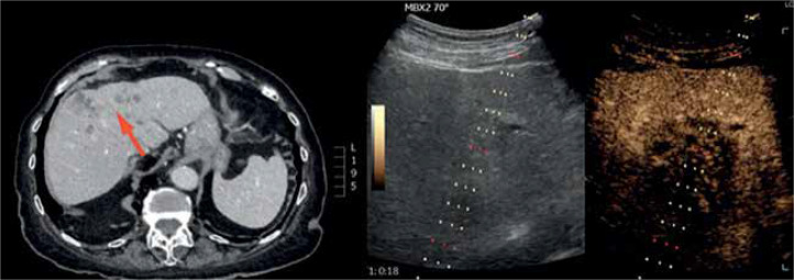
The aim of this study is to determine the usefulness of contrast-enhanced ultrasound (CEUS) in liver biopsy. The popularisation of imaging techniques that visualise the abdominal cavity, especially ultrasonography (USG), has resulted in an increase in the detection of focal liver lesions (FLL). If the results of other imaging modalities (magnetic resonance imaging [MRI] or computed tomography [CT]) are inconclusive, percutaneous liver biopsy should be considered. Taking into account the limitations of using MRI and CT in liver biopsy, this procedure is mostly performed with ultrasound. It is economical, safe, and swift. Whenever it is impossible to visualise lesions in B-mode (a condition necessary for a safe and effective biopsy), it is advisable to use advanced ultrasound techniques - CEUS or fusion imaging. Limitations of fusion imaging include prolonged time of data processing and difficulties in achieving optimal overlap of images. Conversely, CEUS enhances lesion visualisation but is devoid of the mentioned limitations - it is rapid and requires no additional processing. Furthermore, considering the potential of CEUS in the visualisation of focal liver lesions and differentiation of necrotic areas, accompanied by the ability to detect neuroendocrine tumours or its metastasis, we strongly believe that biopsy procedures - especially core needle biopsies - with CEUS assistance are potent tools in contemporary diagnostics. In this paper we want to share the experience of our centre and review the available literature on performing liver biopsies under CEUS guidance.

 求助内容:
求助内容: 应助结果提醒方式:


