{"title":"采用基于二维超声成像的乳腺癌瘤内和瘤周放射组学来确定预测KI-67表达的最佳瘤周范围。","authors":"Wangxing Huang, Songming Zheng, Xiaoyan Zhang, Lina Qi, Min Li, Qinghua Zhang, Zhen Zhen, Xiuwei Yang, Changqin Kong, Dong Li, Guoyong Hua","doi":"10.1007/s40477-025-01049-0","DOIUrl":null,"url":null,"abstract":"<p><strong>Objectives: </strong>Currently, radiomics focuses on intratumoral regions and fixed peritumoral regions, and lacks an optimal peritumoral region taken to predict KI-67 expression. The aim of this study was to develop a machine learning model to analyze ultrasound radiomics features with different regions of peri-tumor fetch values to determine the optimal peri-tumor region for predicting KI-67 expression.</p><p><strong>Methods: </strong>A total of 453 breast cancer patients were included. They were randomly assigned to training and validation sets in a 7:3 ratio. In the training cohort, machine learning models were constructed for intra-tumor and different peri-tumor regions (2 mm, 4 mm, 6 mm, 8 mm, 10 mm), identifying the relevant Ki-67 features for each ROI and comparing the different models to determine the best model. These models were validated using a test cohort to find the most accurate peri-tumor region for Ki-67 prediction. The area under the receiver operating characteristic curve (AUC) was used to evaluate the performance of predicting KI-67 expression, and the Delong test was used to assess the difference between each AUC.SHAP (Shapley Additive Decomposition) was performed to analyze the optimal prediction model and quantify the contribution of major radiomics features.</p><p><strong>Results: </strong>In the validation cohort, the SVM model with the combination of intratumoral and peritumoral 6 mm regions showed the highest prediction effect, with an AUC of 0.9342.The intratumoral and peritumoral 6-mm SVM models showed statistically significant differences (P < 0.05) compared to the other models. SHAP analysis showed that peri-tumoral 6 mm features were more important than intratumoral features.</p><p><strong>Conclusion: </strong>SVM models using intratumoral and peritumoral 6 mm regions showed the best results in prediction of KI-67 expression.</p>","PeriodicalId":51528,"journal":{"name":"Journal of Ultrasound","volume":" ","pages":"709-717"},"PeriodicalIF":1.4000,"publicationDate":"2025-09-01","publicationTypes":"Journal Article","fieldsOfStudy":null,"isOpenAccess":false,"openAccessPdf":"https://www.ncbi.nlm.nih.gov/pmc/articles/PMC12496391/pdf/","citationCount":"0","resultStr":"{\"title\":\"Intratumoral and peritumoral radiomics based on 2D ultrasound imaging in breast cancer was used to determine the optimal peritumoral range for predicting KI-67 expression.\",\"authors\":\"Wangxing Huang, Songming Zheng, Xiaoyan Zhang, Lina Qi, Min Li, Qinghua Zhang, Zhen Zhen, Xiuwei Yang, Changqin Kong, Dong Li, Guoyong Hua\",\"doi\":\"10.1007/s40477-025-01049-0\",\"DOIUrl\":null,\"url\":null,\"abstract\":\"<p><strong>Objectives: </strong>Currently, radiomics focuses on intratumoral regions and fixed peritumoral regions, and lacks an optimal peritumoral region taken to predict KI-67 expression. The aim of this study was to develop a machine learning model to analyze ultrasound radiomics features with different regions of peri-tumor fetch values to determine the optimal peri-tumor region for predicting KI-67 expression.</p><p><strong>Methods: </strong>A total of 453 breast cancer patients were included. They were randomly assigned to training and validation sets in a 7:3 ratio. In the training cohort, machine learning models were constructed for intra-tumor and different peri-tumor regions (2 mm, 4 mm, 6 mm, 8 mm, 10 mm), identifying the relevant Ki-67 features for each ROI and comparing the different models to determine the best model. These models were validated using a test cohort to find the most accurate peri-tumor region for Ki-67 prediction. The area under the receiver operating characteristic curve (AUC) was used to evaluate the performance of predicting KI-67 expression, and the Delong test was used to assess the difference between each AUC.SHAP (Shapley Additive Decomposition) was performed to analyze the optimal prediction model and quantify the contribution of major radiomics features.</p><p><strong>Results: </strong>In the validation cohort, the SVM model with the combination of intratumoral and peritumoral 6 mm regions showed the highest prediction effect, with an AUC of 0.9342.The intratumoral and peritumoral 6-mm SVM models showed statistically significant differences (P < 0.05) compared to the other models. SHAP analysis showed that peri-tumoral 6 mm features were more important than intratumoral features.</p><p><strong>Conclusion: </strong>SVM models using intratumoral and peritumoral 6 mm regions showed the best results in prediction of KI-67 expression.</p>\",\"PeriodicalId\":51528,\"journal\":{\"name\":\"Journal of Ultrasound\",\"volume\":\" \",\"pages\":\"709-717\"},\"PeriodicalIF\":1.4000,\"publicationDate\":\"2025-09-01\",\"publicationTypes\":\"Journal Article\",\"fieldsOfStudy\":null,\"isOpenAccess\":false,\"openAccessPdf\":\"https://www.ncbi.nlm.nih.gov/pmc/articles/PMC12496391/pdf/\",\"citationCount\":\"0\",\"resultStr\":null,\"platform\":\"Semanticscholar\",\"paperid\":null,\"PeriodicalName\":\"Journal of Ultrasound\",\"FirstCategoryId\":\"1085\",\"ListUrlMain\":\"https://doi.org/10.1007/s40477-025-01049-0\",\"RegionNum\":0,\"RegionCategory\":null,\"ArticlePicture\":[],\"TitleCN\":null,\"AbstractTextCN\":null,\"PMCID\":null,\"EPubDate\":\"2025/7/10 0:00:00\",\"PubModel\":\"Epub\",\"JCR\":\"Q3\",\"JCRName\":\"RADIOLOGY, NUCLEAR MEDICINE & MEDICAL IMAGING\",\"Score\":null,\"Total\":0}","platform":"Semanticscholar","paperid":null,"PeriodicalName":"Journal of Ultrasound","FirstCategoryId":"1085","ListUrlMain":"https://doi.org/10.1007/s40477-025-01049-0","RegionNum":0,"RegionCategory":null,"ArticlePicture":[],"TitleCN":null,"AbstractTextCN":null,"PMCID":null,"EPubDate":"2025/7/10 0:00:00","PubModel":"Epub","JCR":"Q3","JCRName":"RADIOLOGY, NUCLEAR MEDICINE & MEDICAL IMAGING","Score":null,"Total":0}
Intratumoral and peritumoral radiomics based on 2D ultrasound imaging in breast cancer was used to determine the optimal peritumoral range for predicting KI-67 expression.
Objectives: Currently, radiomics focuses on intratumoral regions and fixed peritumoral regions, and lacks an optimal peritumoral region taken to predict KI-67 expression. The aim of this study was to develop a machine learning model to analyze ultrasound radiomics features with different regions of peri-tumor fetch values to determine the optimal peri-tumor region for predicting KI-67 expression.
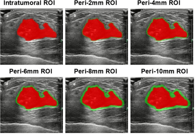
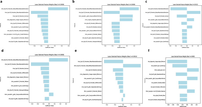
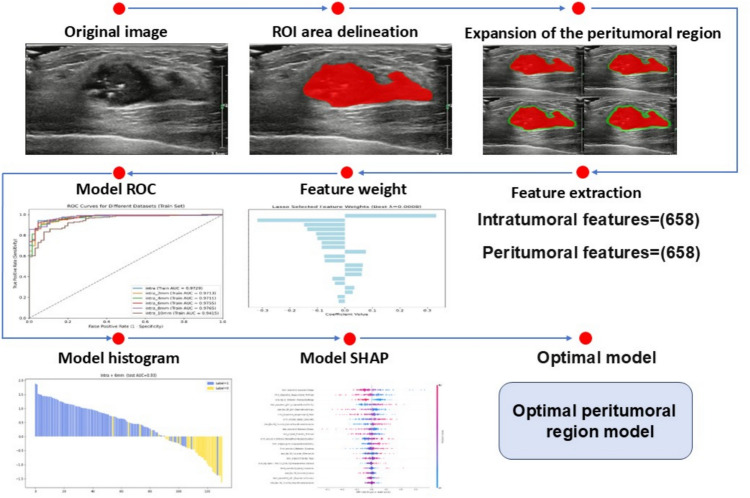
Methods: A total of 453 breast cancer patients were included. They were randomly assigned to training and validation sets in a 7:3 ratio. In the training cohort, machine learning models were constructed for intra-tumor and different peri-tumor regions (2 mm, 4 mm, 6 mm, 8 mm, 10 mm), identifying the relevant Ki-67 features for each ROI and comparing the different models to determine the best model. These models were validated using a test cohort to find the most accurate peri-tumor region for Ki-67 prediction. The area under the receiver operating characteristic curve (AUC) was used to evaluate the performance of predicting KI-67 expression, and the Delong test was used to assess the difference between each AUC.SHAP (Shapley Additive Decomposition) was performed to analyze the optimal prediction model and quantify the contribution of major radiomics features.
Results: In the validation cohort, the SVM model with the combination of intratumoral and peritumoral 6 mm regions showed the highest prediction effect, with an AUC of 0.9342.The intratumoral and peritumoral 6-mm SVM models showed statistically significant differences (P < 0.05) compared to the other models. SHAP analysis showed that peri-tumoral 6 mm features were more important than intratumoral features.
Conclusion: SVM models using intratumoral and peritumoral 6 mm regions showed the best results in prediction of KI-67 expression.
期刊介绍:
The Journal of Ultrasound is the official journal of the Italian Society for Ultrasound in Medicine and Biology (SIUMB). The journal publishes original contributions (research and review articles, case reports, technical reports and letters to the editor) on significant advances in clinical diagnostic, interventional and therapeutic applications, clinical techniques, the physics, engineering and technology of ultrasound in medicine and biology, and in cross-sectional diagnostic imaging. The official language of Journal of Ultrasound is English.

 求助内容:
求助内容: 应助结果提醒方式:


