{"title":"上颌窦内有一个大的含牙囊肿并伴有上颌第三磨牙异位的罕见病例报告。","authors":"Marika Ramishvili, Leila Atskvereli, Giorgi Menabde, Marika Zurmukhtashvili, Sopio Samkharadze, Giorgi Dugashvili, Luc Marks","doi":"10.1155/crid/2436615","DOIUrl":null,"url":null,"abstract":"<p><p>Ectopic eruption of permanent molars is an uncommon developmental anomaly characterized by abnormal tooth positioning, which can lead to significant complications. In rare instances, ectopic molars may be associated with dentigerous cysts, particularly within the maxillary sinus, posing challenges for diagnosis and management. This report discusses a rare case of a 58-year-old male who presented with chronic right maxillary sinusitis, intermittent facial pain, and purulent nasal and oral discharge. Radiological evaluation, including cone beam computed tomography (CBCT), revealed a completely opacified right maxillary sinus containing an ectopic maxillary molar. Additionally, a large cystic lesion consistent with a dentigerous cyst was found, occupying the entire sinus cavity. Surgical management was performed using the Caldwell-Luc approach under general anesthesia. This involved creating a bone window in the anterior maxillary wall to facilitate the removal of the ectopic tooth and the associated cystic lesion. Histopathological examination confirmed the presence of a dentigerous cyst exhibiting chronic inflammatory infiltration and fibrosis. Ectopic molars in the maxillary sinus are often asymptomatic but can present with recurrent sinusitis, pain, and oroantral communication. The existence of a large dentigerous cyst heightens the risk of complications and may obscure radiological interpretation due to sinus opacification. This case highlights the necessity of comprehensive imaging and early surgical intervention to prevent long-term complications. Awareness of such rare conditions can help clinicians in prompt diagnosis and appropriate management, ultimately preserving sinus function and minimizing further issues.</p>","PeriodicalId":46841,"journal":{"name":"Case Reports in Dentistry","volume":"2025 ","pages":"2436615"},"PeriodicalIF":0.9000,"publicationDate":"2025-07-03","publicationTypes":"Journal Article","fieldsOfStudy":null,"isOpenAccess":false,"openAccessPdf":"https://www.ncbi.nlm.nih.gov/pmc/articles/PMC12245510/pdf/","citationCount":"0","resultStr":"{\"title\":\"A Rare Case Report of a Large Dentigerous Cyst in the Maxillary Sinus Associated With an Ectopic Maxillary Third Molar.\",\"authors\":\"Marika Ramishvili, Leila Atskvereli, Giorgi Menabde, Marika Zurmukhtashvili, Sopio Samkharadze, Giorgi Dugashvili, Luc Marks\",\"doi\":\"10.1155/crid/2436615\",\"DOIUrl\":null,\"url\":null,\"abstract\":\"<p><p>Ectopic eruption of permanent molars is an uncommon developmental anomaly characterized by abnormal tooth positioning, which can lead to significant complications. In rare instances, ectopic molars may be associated with dentigerous cysts, particularly within the maxillary sinus, posing challenges for diagnosis and management. This report discusses a rare case of a 58-year-old male who presented with chronic right maxillary sinusitis, intermittent facial pain, and purulent nasal and oral discharge. Radiological evaluation, including cone beam computed tomography (CBCT), revealed a completely opacified right maxillary sinus containing an ectopic maxillary molar. Additionally, a large cystic lesion consistent with a dentigerous cyst was found, occupying the entire sinus cavity. Surgical management was performed using the Caldwell-Luc approach under general anesthesia. This involved creating a bone window in the anterior maxillary wall to facilitate the removal of the ectopic tooth and the associated cystic lesion. Histopathological examination confirmed the presence of a dentigerous cyst exhibiting chronic inflammatory infiltration and fibrosis. Ectopic molars in the maxillary sinus are often asymptomatic but can present with recurrent sinusitis, pain, and oroantral communication. The existence of a large dentigerous cyst heightens the risk of complications and may obscure radiological interpretation due to sinus opacification. This case highlights the necessity of comprehensive imaging and early surgical intervention to prevent long-term complications. Awareness of such rare conditions can help clinicians in prompt diagnosis and appropriate management, ultimately preserving sinus function and minimizing further issues.</p>\",\"PeriodicalId\":46841,\"journal\":{\"name\":\"Case Reports in Dentistry\",\"volume\":\"2025 \",\"pages\":\"2436615\"},\"PeriodicalIF\":0.9000,\"publicationDate\":\"2025-07-03\",\"publicationTypes\":\"Journal Article\",\"fieldsOfStudy\":null,\"isOpenAccess\":false,\"openAccessPdf\":\"https://www.ncbi.nlm.nih.gov/pmc/articles/PMC12245510/pdf/\",\"citationCount\":\"0\",\"resultStr\":null,\"platform\":\"Semanticscholar\",\"paperid\":null,\"PeriodicalName\":\"Case Reports in Dentistry\",\"FirstCategoryId\":\"1085\",\"ListUrlMain\":\"https://doi.org/10.1155/crid/2436615\",\"RegionNum\":0,\"RegionCategory\":null,\"ArticlePicture\":[],\"TitleCN\":null,\"AbstractTextCN\":null,\"PMCID\":null,\"EPubDate\":\"2025/1/1 0:00:00\",\"PubModel\":\"eCollection\",\"JCR\":\"Q4\",\"JCRName\":\"DENTISTRY, ORAL SURGERY & MEDICINE\",\"Score\":null,\"Total\":0}","platform":"Semanticscholar","paperid":null,"PeriodicalName":"Case Reports in Dentistry","FirstCategoryId":"1085","ListUrlMain":"https://doi.org/10.1155/crid/2436615","RegionNum":0,"RegionCategory":null,"ArticlePicture":[],"TitleCN":null,"AbstractTextCN":null,"PMCID":null,"EPubDate":"2025/1/1 0:00:00","PubModel":"eCollection","JCR":"Q4","JCRName":"DENTISTRY, ORAL SURGERY & MEDICINE","Score":null,"Total":0}
A Rare Case Report of a Large Dentigerous Cyst in the Maxillary Sinus Associated With an Ectopic Maxillary Third Molar.
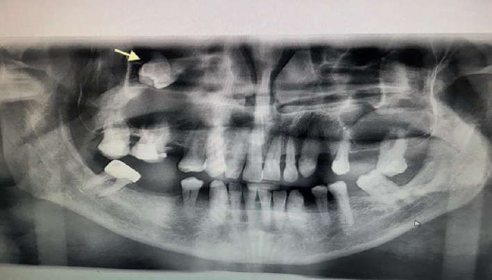
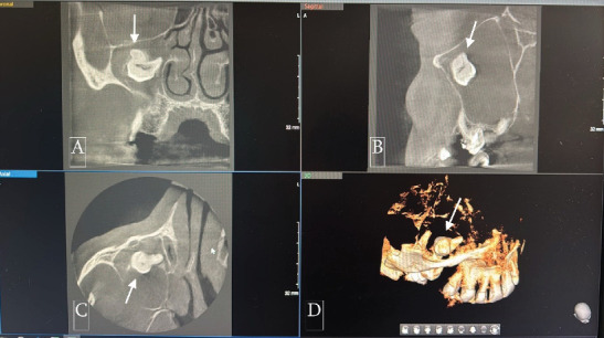
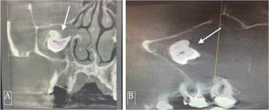
Ectopic eruption of permanent molars is an uncommon developmental anomaly characterized by abnormal tooth positioning, which can lead to significant complications. In rare instances, ectopic molars may be associated with dentigerous cysts, particularly within the maxillary sinus, posing challenges for diagnosis and management. This report discusses a rare case of a 58-year-old male who presented with chronic right maxillary sinusitis, intermittent facial pain, and purulent nasal and oral discharge. Radiological evaluation, including cone beam computed tomography (CBCT), revealed a completely opacified right maxillary sinus containing an ectopic maxillary molar. Additionally, a large cystic lesion consistent with a dentigerous cyst was found, occupying the entire sinus cavity. Surgical management was performed using the Caldwell-Luc approach under general anesthesia. This involved creating a bone window in the anterior maxillary wall to facilitate the removal of the ectopic tooth and the associated cystic lesion. Histopathological examination confirmed the presence of a dentigerous cyst exhibiting chronic inflammatory infiltration and fibrosis. Ectopic molars in the maxillary sinus are often asymptomatic but can present with recurrent sinusitis, pain, and oroantral communication. The existence of a large dentigerous cyst heightens the risk of complications and may obscure radiological interpretation due to sinus opacification. This case highlights the necessity of comprehensive imaging and early surgical intervention to prevent long-term complications. Awareness of such rare conditions can help clinicians in prompt diagnosis and appropriate management, ultimately preserving sinus function and minimizing further issues.
期刊介绍:
Case Reports in Dentistry is a peer-reviewed, Open Access journal that publishes case reports and case series in all areas of dentistry, including periodontal diseases, dental implants, oral pathology, as well as oral and maxillofacial surgery.

 求助内容:
求助内容: 应助结果提醒方式:


