Pedro Beredjiklian, Brianna Fram, Jason Core, Joshua I Ng, Robert Pugliese, Stephanie A Kwan, Michael Rivlin
{"title":"肩关节周围骨折和肘关节周围骨折的3D打印模型改善骨科学员的手术决策。","authors":"Pedro Beredjiklian, Brianna Fram, Jason Core, Joshua I Ng, Robert Pugliese, Stephanie A Kwan, Michael Rivlin","doi":"10.22038/ABJS.2024.81232.3707","DOIUrl":null,"url":null,"abstract":"<p><strong>Objectives: </strong>Periarticular fractures of the shoulder and elbow are spatially complex injuries that may be challenging to interpret on radiographs and advanced imaging. As three-dimensional (3D) printing technology has become less expensive and more available, 3D printed fracture models have gained attention for use in surgical preparation. In this study, we evaluated the effects of 3D printed fracture models on orthopedic trainee surgical planning and injury understanding for injuries of the shoulder and elbow.</p><p><strong>Methods: </strong>Models of periarticular fractures of the shoulder and elbow were manufactured by 3D printing at the medical school design lab. Eleven Orthopedic trainees viewed X-rays and computed tomography (CT) scans for each injury, and completed a preoperative questionnaires. They were then given access to the 3D model of each injury, in addition to the previously viewed imaging. They again completed a preoperative plan and questionnaire. Preoperative plans were graded for feasibility by a preestablished template. Results were compared for each participant with and without the 3D models.</p><p><strong>Results: </strong>Within all trainees and fractures, trainees were more likely to have feasible preoperative plans when given a 3D model, compared to access to x-rays and CT scans alone (74% vs. 62%). In all cases where preoperative plans were changed after handling the 3D models (46/77 changed, 60%), they stayed static or improved in feasibility. Participants reported significantly improved understanding of injury anatomy (P<0.0001), increased confidence in choosing operative positioning and surgical approaches (P<0.0001), desired implants (P=0.011), and better conceptualization of how to perform fracture reduction (P=0.0038).</p><p><strong>Conclusion: </strong>Orthopedic trainees benefit from 3D printed fracture models when performing preoperative planning of complex periarticular shoulder and elbow injuries. Given the rarity and difficulty of these injuries, use of this technology could allow for shortened learning curves and improved surgical results in the field of orthopedic fracture care.</p>","PeriodicalId":46704,"journal":{"name":"Archives of Bone and Joint Surgery-ABJS","volume":"13 6","pages":"337-344"},"PeriodicalIF":1.8000,"publicationDate":"2025-01-01","publicationTypes":"Journal Article","fieldsOfStudy":null,"isOpenAccess":false,"openAccessPdf":"https://www.ncbi.nlm.nih.gov/pmc/articles/PMC12238857/pdf/","citationCount":"0","resultStr":"{\"title\":\"3D Printed Models of Periarticular Fractures of the Shoulder and Elbow Improve Surgical Decision Making in Orthopedic Trainees.\",\"authors\":\"Pedro Beredjiklian, Brianna Fram, Jason Core, Joshua I Ng, Robert Pugliese, Stephanie A Kwan, Michael Rivlin\",\"doi\":\"10.22038/ABJS.2024.81232.3707\",\"DOIUrl\":null,\"url\":null,\"abstract\":\"<p><strong>Objectives: </strong>Periarticular fractures of the shoulder and elbow are spatially complex injuries that may be challenging to interpret on radiographs and advanced imaging. As three-dimensional (3D) printing technology has become less expensive and more available, 3D printed fracture models have gained attention for use in surgical preparation. In this study, we evaluated the effects of 3D printed fracture models on orthopedic trainee surgical planning and injury understanding for injuries of the shoulder and elbow.</p><p><strong>Methods: </strong>Models of periarticular fractures of the shoulder and elbow were manufactured by 3D printing at the medical school design lab. Eleven Orthopedic trainees viewed X-rays and computed tomography (CT) scans for each injury, and completed a preoperative questionnaires. They were then given access to the 3D model of each injury, in addition to the previously viewed imaging. They again completed a preoperative plan and questionnaire. Preoperative plans were graded for feasibility by a preestablished template. Results were compared for each participant with and without the 3D models.</p><p><strong>Results: </strong>Within all trainees and fractures, trainees were more likely to have feasible preoperative plans when given a 3D model, compared to access to x-rays and CT scans alone (74% vs. 62%). In all cases where preoperative plans were changed after handling the 3D models (46/77 changed, 60%), they stayed static or improved in feasibility. Participants reported significantly improved understanding of injury anatomy (P<0.0001), increased confidence in choosing operative positioning and surgical approaches (P<0.0001), desired implants (P=0.011), and better conceptualization of how to perform fracture reduction (P=0.0038).</p><p><strong>Conclusion: </strong>Orthopedic trainees benefit from 3D printed fracture models when performing preoperative planning of complex periarticular shoulder and elbow injuries. Given the rarity and difficulty of these injuries, use of this technology could allow for shortened learning curves and improved surgical results in the field of orthopedic fracture care.</p>\",\"PeriodicalId\":46704,\"journal\":{\"name\":\"Archives of Bone and Joint Surgery-ABJS\",\"volume\":\"13 6\",\"pages\":\"337-344\"},\"PeriodicalIF\":1.8000,\"publicationDate\":\"2025-01-01\",\"publicationTypes\":\"Journal Article\",\"fieldsOfStudy\":null,\"isOpenAccess\":false,\"openAccessPdf\":\"https://www.ncbi.nlm.nih.gov/pmc/articles/PMC12238857/pdf/\",\"citationCount\":\"0\",\"resultStr\":null,\"platform\":\"Semanticscholar\",\"paperid\":null,\"PeriodicalName\":\"Archives of Bone and Joint Surgery-ABJS\",\"FirstCategoryId\":\"1085\",\"ListUrlMain\":\"https://doi.org/10.22038/ABJS.2024.81232.3707\",\"RegionNum\":0,\"RegionCategory\":null,\"ArticlePicture\":[],\"TitleCN\":null,\"AbstractTextCN\":null,\"PMCID\":null,\"EPubDate\":\"\",\"PubModel\":\"\",\"JCR\":\"Q3\",\"JCRName\":\"ORTHOPEDICS\",\"Score\":null,\"Total\":0}","platform":"Semanticscholar","paperid":null,"PeriodicalName":"Archives of Bone and Joint Surgery-ABJS","FirstCategoryId":"1085","ListUrlMain":"https://doi.org/10.22038/ABJS.2024.81232.3707","RegionNum":0,"RegionCategory":null,"ArticlePicture":[],"TitleCN":null,"AbstractTextCN":null,"PMCID":null,"EPubDate":"","PubModel":"","JCR":"Q3","JCRName":"ORTHOPEDICS","Score":null,"Total":0}
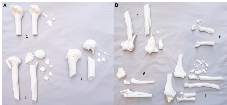
3D Printed Models of Periarticular Fractures of the Shoulder and Elbow Improve Surgical Decision Making in Orthopedic Trainees.
Objectives: Periarticular fractures of the shoulder and elbow are spatially complex injuries that may be challenging to interpret on radiographs and advanced imaging. As three-dimensional (3D) printing technology has become less expensive and more available, 3D printed fracture models have gained attention for use in surgical preparation. In this study, we evaluated the effects of 3D printed fracture models on orthopedic trainee surgical planning and injury understanding for injuries of the shoulder and elbow.
Methods: Models of periarticular fractures of the shoulder and elbow were manufactured by 3D printing at the medical school design lab. Eleven Orthopedic trainees viewed X-rays and computed tomography (CT) scans for each injury, and completed a preoperative questionnaires. They were then given access to the 3D model of each injury, in addition to the previously viewed imaging. They again completed a preoperative plan and questionnaire. Preoperative plans were graded for feasibility by a preestablished template. Results were compared for each participant with and without the 3D models.
Results: Within all trainees and fractures, trainees were more likely to have feasible preoperative plans when given a 3D model, compared to access to x-rays and CT scans alone (74% vs. 62%). In all cases where preoperative plans were changed after handling the 3D models (46/77 changed, 60%), they stayed static or improved in feasibility. Participants reported significantly improved understanding of injury anatomy (P<0.0001), increased confidence in choosing operative positioning and surgical approaches (P<0.0001), desired implants (P=0.011), and better conceptualization of how to perform fracture reduction (P=0.0038).
Conclusion: Orthopedic trainees benefit from 3D printed fracture models when performing preoperative planning of complex periarticular shoulder and elbow injuries. Given the rarity and difficulty of these injuries, use of this technology could allow for shortened learning curves and improved surgical results in the field of orthopedic fracture care.
期刊介绍:
The Archives of Bone and Joint Surgery (ABJS) aims to encourage a better understanding of all aspects of Orthopedic Sciences. The journal accepts scientific papers including original research, review article, short communication, case report, and letter to the editor in all fields of bone, joint, musculoskeletal surgery and related researches. The Archives of Bone and Joint Surgery (ABJS) will publish papers in all aspects of today`s modern orthopedic sciences including: Arthroscopy, Arthroplasty, Sport Medicine, Reconstruction, Hand and Upper Extremity, Pediatric Orthopedics, Spine, Trauma, Foot and Ankle, Tumor, Joint Rheumatic Disease, Skeletal Imaging, Orthopedic Physical Therapy, Rehabilitation, Orthopedic Basic Sciences (Biomechanics, Biotechnology, Biomaterial..).

 求助内容:
求助内容: 应助结果提醒方式:


