Nilüfer Bıçakcı, Fatih Batı, Güler Silov, Banu Kırtıloğlu
{"title":"在肿瘤学PET/CT成像中偶然发现背弹性纤维瘤:一项回顾性单中心分析。","authors":"Nilüfer Bıçakcı, Fatih Batı, Güler Silov, Banu Kırtıloğlu","doi":"10.47717/turkjsurg.2025.2025-5-18","DOIUrl":null,"url":null,"abstract":"<p><strong>Objective: </strong>To evaluate the morphological and metabolic characteristics of incidentally detected elastofibroma dorsi (EFD) on F-18 florodeoksiglukoz (FDG) positron emission tomography/computed tomography (PET/CT) and their longitudinal changes in oncologic patients.</p><p><strong>Material and methods: </strong>We retrospectively reviewed 42 197 PET/CT scans performed at our institution between January 2019 and September 2023. EFD was incidentally identified in 20 patients (0.05%). Patient demographics, primary malignancy, lesion localization, dimensions, and maximum standardized uptake values (SUV<sub>max</sub>) were recorded. Measurements were obtained before treatment and at the next 3‑month follow‑up. Statistical analyses included Mann‑Whitney U, Shapiro-Wilk and Spearman correlation tests; significance was set at p<0.05.</p><p><strong>Results: </strong>The cohort comprised 17 females (85%) and 3 males (15%) with a median age of 67 years (range, 47-83). Primary diagnoses were breast cancer (n=8, 40%) and various other malignancies (n=12, 60%). Lesions were bilateral in 75% of cases. Pre‑treatment lesion size ranged from 10 to 55 mm; median SUV<sub>max</sub> was 2.4 (right) and 2.5 (left). No significant differences in baseline size or SUV<sub>max</sub> were observed between breast and other cancers. A moderate correlation existed between right and left SUV<sub>max</sub> (r=0.641; p=0.010). After 3 months, only the left longest diameter showed a statistically significant decrease (median, 45.0 mm vs. 43.0 mm; p=0.034), which may reflect measurement variability or positional factors rather than true biological change. SUV<sub>max</sub> values remained stable.</p><p><strong>Conclusion: </strong>Incidentally detected EFD on PET/CT exhibits low to moderate and stable FDG uptake and predominantly bilateral localization. Recognition of its characteristic features can prevent unnecessary interventions.</p>","PeriodicalId":23374,"journal":{"name":"Turkish Journal of Surgery","volume":" ","pages":"313-320"},"PeriodicalIF":0.6000,"publicationDate":"2025-09-03","publicationTypes":"Journal Article","fieldsOfStudy":null,"isOpenAccess":false,"openAccessPdf":"https://www.ncbi.nlm.nih.gov/pmc/articles/PMC12406630/pdf/","citationCount":"0","resultStr":"{\"title\":\"Incidental identification of elastofibroma dorsi in oncologic PET/CT imaging: a retrospective single-center analysis.\",\"authors\":\"Nilüfer Bıçakcı, Fatih Batı, Güler Silov, Banu Kırtıloğlu\",\"doi\":\"10.47717/turkjsurg.2025.2025-5-18\",\"DOIUrl\":null,\"url\":null,\"abstract\":\"<p><strong>Objective: </strong>To evaluate the morphological and metabolic characteristics of incidentally detected elastofibroma dorsi (EFD) on F-18 florodeoksiglukoz (FDG) positron emission tomography/computed tomography (PET/CT) and their longitudinal changes in oncologic patients.</p><p><strong>Material and methods: </strong>We retrospectively reviewed 42 197 PET/CT scans performed at our institution between January 2019 and September 2023. EFD was incidentally identified in 20 patients (0.05%). Patient demographics, primary malignancy, lesion localization, dimensions, and maximum standardized uptake values (SUV<sub>max</sub>) were recorded. Measurements were obtained before treatment and at the next 3‑month follow‑up. Statistical analyses included Mann‑Whitney U, Shapiro-Wilk and Spearman correlation tests; significance was set at p<0.05.</p><p><strong>Results: </strong>The cohort comprised 17 females (85%) and 3 males (15%) with a median age of 67 years (range, 47-83). Primary diagnoses were breast cancer (n=8, 40%) and various other malignancies (n=12, 60%). Lesions were bilateral in 75% of cases. Pre‑treatment lesion size ranged from 10 to 55 mm; median SUV<sub>max</sub> was 2.4 (right) and 2.5 (left). No significant differences in baseline size or SUV<sub>max</sub> were observed between breast and other cancers. A moderate correlation existed between right and left SUV<sub>max</sub> (r=0.641; p=0.010). After 3 months, only the left longest diameter showed a statistically significant decrease (median, 45.0 mm vs. 43.0 mm; p=0.034), which may reflect measurement variability or positional factors rather than true biological change. SUV<sub>max</sub> values remained stable.</p><p><strong>Conclusion: </strong>Incidentally detected EFD on PET/CT exhibits low to moderate and stable FDG uptake and predominantly bilateral localization. Recognition of its characteristic features can prevent unnecessary interventions.</p>\",\"PeriodicalId\":23374,\"journal\":{\"name\":\"Turkish Journal of Surgery\",\"volume\":\" \",\"pages\":\"313-320\"},\"PeriodicalIF\":0.6000,\"publicationDate\":\"2025-09-03\",\"publicationTypes\":\"Journal Article\",\"fieldsOfStudy\":null,\"isOpenAccess\":false,\"openAccessPdf\":\"https://www.ncbi.nlm.nih.gov/pmc/articles/PMC12406630/pdf/\",\"citationCount\":\"0\",\"resultStr\":null,\"platform\":\"Semanticscholar\",\"paperid\":null,\"PeriodicalName\":\"Turkish Journal of Surgery\",\"FirstCategoryId\":\"1085\",\"ListUrlMain\":\"https://doi.org/10.47717/turkjsurg.2025.2025-5-18\",\"RegionNum\":0,\"RegionCategory\":null,\"ArticlePicture\":[],\"TitleCN\":null,\"AbstractTextCN\":null,\"PMCID\":null,\"EPubDate\":\"2025/7/11 0:00:00\",\"PubModel\":\"Epub\",\"JCR\":\"Q4\",\"JCRName\":\"SURGERY\",\"Score\":null,\"Total\":0}","platform":"Semanticscholar","paperid":null,"PeriodicalName":"Turkish Journal of Surgery","FirstCategoryId":"1085","ListUrlMain":"https://doi.org/10.47717/turkjsurg.2025.2025-5-18","RegionNum":0,"RegionCategory":null,"ArticlePicture":[],"TitleCN":null,"AbstractTextCN":null,"PMCID":null,"EPubDate":"2025/7/11 0:00:00","PubModel":"Epub","JCR":"Q4","JCRName":"SURGERY","Score":null,"Total":0}
引用次数: 0
摘要
目的:评价F-18 florodeoksiglukoz (FDG)正电子发射断层扫描/计算机断层扫描(PET/CT)偶然发现的背弹性纤维瘤(EFD)的形态学和代谢特征及其在肿瘤患者中的纵向变化。材料和方法:我们回顾性回顾了2019年1月至2023年9月期间在我院进行的42 197次PET/CT扫描。20例患者偶然发现EFD(0.05%)。记录患者人口统计学、原发恶性、病变定位、尺寸和最大标准化摄取值(SUVmax)。在治疗前和接下来的3个月随访时进行测量。统计分析包括Mann - Whitney U、Shapiro-Wilk和Spearman相关检验;结果:该队列包括17名女性(85%)和3名男性(15%),中位年龄为67岁(范围47-83)。最初诊断为乳腺癌(n=8, 40%)和各种其他恶性肿瘤(n=12, 60%)。75%的病例为双侧病变。治疗前病变大小为10 ~ 55 mm;中位SUVmax分别为2.4(右)和2.5(左)。在乳腺癌和其他癌症之间没有观察到基线大小或SUVmax的显著差异。左、右SUVmax存在中度相关(r=0.641;p = 0.010)。3个月后,只有左最长直径有统计学意义的减少(中位数,45.0 mm vs. 43.0 mm;P =0.034),这可能反映了测量变异性或位置因素,而不是真正的生物学变化。SUVmax值保持稳定。结论:偶然发现的EFD在PET/CT上表现为低至中度和稳定的FDG摄取,主要是双侧定位。认识到它的特征可以防止不必要的干预。
Incidental identification of elastofibroma dorsi in oncologic PET/CT imaging: a retrospective single-center analysis.
Objective: To evaluate the morphological and metabolic characteristics of incidentally detected elastofibroma dorsi (EFD) on F-18 florodeoksiglukoz (FDG) positron emission tomography/computed tomography (PET/CT) and their longitudinal changes in oncologic patients.
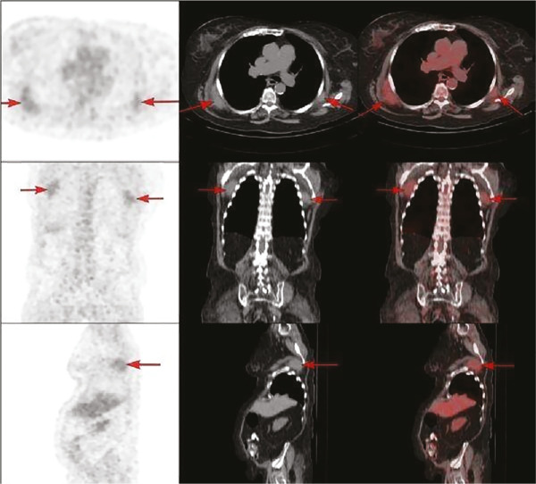
Material and methods: We retrospectively reviewed 42 197 PET/CT scans performed at our institution between January 2019 and September 2023. EFD was incidentally identified in 20 patients (0.05%). Patient demographics, primary malignancy, lesion localization, dimensions, and maximum standardized uptake values (SUVmax) were recorded. Measurements were obtained before treatment and at the next 3‑month follow‑up. Statistical analyses included Mann‑Whitney U, Shapiro-Wilk and Spearman correlation tests; significance was set at p<0.05.
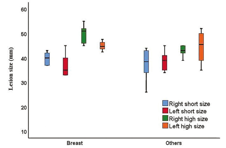
Results: The cohort comprised 17 females (85%) and 3 males (15%) with a median age of 67 years (range, 47-83). Primary diagnoses were breast cancer (n=8, 40%) and various other malignancies (n=12, 60%). Lesions were bilateral in 75% of cases. Pre‑treatment lesion size ranged from 10 to 55 mm; median SUVmax was 2.4 (right) and 2.5 (left). No significant differences in baseline size or SUVmax were observed between breast and other cancers. A moderate correlation existed between right and left SUVmax (r=0.641; p=0.010). After 3 months, only the left longest diameter showed a statistically significant decrease (median, 45.0 mm vs. 43.0 mm; p=0.034), which may reflect measurement variability or positional factors rather than true biological change. SUVmax values remained stable.
Conclusion: Incidentally detected EFD on PET/CT exhibits low to moderate and stable FDG uptake and predominantly bilateral localization. Recognition of its characteristic features can prevent unnecessary interventions.

 求助内容:
求助内容: 应助结果提醒方式:


