Li Fu, Baixu Zhou, Xia Jiang, Jingliang Cheng, Qiang Wu, Junjiang Fu
{"title":"人SPATA3在小鼠睾丸细胞增殖中的作用及SPATA3的表达模式。","authors":"Li Fu, Baixu Zhou, Xia Jiang, Jingliang Cheng, Qiang Wu, Junjiang Fu","doi":"10.3892/mmr.2025.13620","DOIUrl":null,"url":null,"abstract":"<p><p>Male infertility predominantly manifests as abnormal spermatogenesis and maturation, abnormal semen quality, chromosomal abnormalities, and endocrine dysfunction. The present study aimed to explore the role, expression pattern and localization of spermatogenesis‑associated protein 3 (SPATA3) in the testis, and to analyze its spatiotemporal expression and function. Using previously acquired plasmids as templates, recombinant plasmids of different SPATA3 isoforms were constructed. Cell Counting Kit‑8 assay was used to analyze cell proliferation. Reverse transcription‑PCR and western blotting were used to determine the mRNA and protein expression levels, respectively. Different SPATA3 isoforms were induced to be overexpressed, among which SPATA3‑I1 and SPATA3‑I2 promoted cell proliferation. The subcellular localization results indicated that the green fluorescent protein fusion proteins of various isoforms were mainly localized in the nucleus. However, the fluorescence of the fusion proteins pEGFP‑C3‑SPATA3‑I3 and pEGFP‑C3‑SPATA3‑I4 tended to be distributed in the cytoplasm, accompanied by a decrease in nuclear fluorescence. Additionally, the SPATA3‑I2 isoform protein displayed notable tissue‑specific expression in testes. Notably, the SPATA3‑I2 isoform protein was not expressed in embryos or during the early development stage of the mice, yet it was highly expressed in the testes of mice aged 23‑57 days (3‑8 weeks). Immunohistochemistry revealed that the SPATA3‑I2 isoform protein was located mainly in round and elongated spermatids within the spermatogenic epithelial cells. In conclusion, the findings highlighted that SPATA3 isoforms had differential subcellular localizations and that SPATA3‑I2 exhibited a specific spatiotemporal expression pattern, suggesting its association with spermatogenesis and sperm maturation.</p>","PeriodicalId":18818,"journal":{"name":"Molecular medicine reports","volume":"32 3","pages":""},"PeriodicalIF":3.5000,"publicationDate":"2025-09-01","publicationTypes":"Journal Article","fieldsOfStudy":null,"isOpenAccess":false,"openAccessPdf":"https://www.ncbi.nlm.nih.gov/pmc/articles/PMC12284469/pdf/","citationCount":"0","resultStr":"{\"title\":\"Roles of human SPATA3 in cell proliferation and expression pattern of <i>Spata3</i> in mouse testis.\",\"authors\":\"Li Fu, Baixu Zhou, Xia Jiang, Jingliang Cheng, Qiang Wu, Junjiang Fu\",\"doi\":\"10.3892/mmr.2025.13620\",\"DOIUrl\":null,\"url\":null,\"abstract\":\"<p><p>Male infertility predominantly manifests as abnormal spermatogenesis and maturation, abnormal semen quality, chromosomal abnormalities, and endocrine dysfunction. The present study aimed to explore the role, expression pattern and localization of spermatogenesis‑associated protein 3 (SPATA3) in the testis, and to analyze its spatiotemporal expression and function. Using previously acquired plasmids as templates, recombinant plasmids of different SPATA3 isoforms were constructed. Cell Counting Kit‑8 assay was used to analyze cell proliferation. Reverse transcription‑PCR and western blotting were used to determine the mRNA and protein expression levels, respectively. Different SPATA3 isoforms were induced to be overexpressed, among which SPATA3‑I1 and SPATA3‑I2 promoted cell proliferation. The subcellular localization results indicated that the green fluorescent protein fusion proteins of various isoforms were mainly localized in the nucleus. However, the fluorescence of the fusion proteins pEGFP‑C3‑SPATA3‑I3 and pEGFP‑C3‑SPATA3‑I4 tended to be distributed in the cytoplasm, accompanied by a decrease in nuclear fluorescence. Additionally, the SPATA3‑I2 isoform protein displayed notable tissue‑specific expression in testes. Notably, the SPATA3‑I2 isoform protein was not expressed in embryos or during the early development stage of the mice, yet it was highly expressed in the testes of mice aged 23‑57 days (3‑8 weeks). Immunohistochemistry revealed that the SPATA3‑I2 isoform protein was located mainly in round and elongated spermatids within the spermatogenic epithelial cells. In conclusion, the findings highlighted that SPATA3 isoforms had differential subcellular localizations and that SPATA3‑I2 exhibited a specific spatiotemporal expression pattern, suggesting its association with spermatogenesis and sperm maturation.</p>\",\"PeriodicalId\":18818,\"journal\":{\"name\":\"Molecular medicine reports\",\"volume\":\"32 3\",\"pages\":\"\"},\"PeriodicalIF\":3.5000,\"publicationDate\":\"2025-09-01\",\"publicationTypes\":\"Journal Article\",\"fieldsOfStudy\":null,\"isOpenAccess\":false,\"openAccessPdf\":\"https://www.ncbi.nlm.nih.gov/pmc/articles/PMC12284469/pdf/\",\"citationCount\":\"0\",\"resultStr\":null,\"platform\":\"Semanticscholar\",\"paperid\":null,\"PeriodicalName\":\"Molecular medicine reports\",\"FirstCategoryId\":\"3\",\"ListUrlMain\":\"https://doi.org/10.3892/mmr.2025.13620\",\"RegionNum\":3,\"RegionCategory\":\"医学\",\"ArticlePicture\":[],\"TitleCN\":null,\"AbstractTextCN\":null,\"PMCID\":null,\"EPubDate\":\"2025/7/11 0:00:00\",\"PubModel\":\"Epub\",\"JCR\":\"Q2\",\"JCRName\":\"MEDICINE, RESEARCH & EXPERIMENTAL\",\"Score\":null,\"Total\":0}","platform":"Semanticscholar","paperid":null,"PeriodicalName":"Molecular medicine reports","FirstCategoryId":"3","ListUrlMain":"https://doi.org/10.3892/mmr.2025.13620","RegionNum":3,"RegionCategory":"医学","ArticlePicture":[],"TitleCN":null,"AbstractTextCN":null,"PMCID":null,"EPubDate":"2025/7/11 0:00:00","PubModel":"Epub","JCR":"Q2","JCRName":"MEDICINE, RESEARCH & EXPERIMENTAL","Score":null,"Total":0}
Roles of human SPATA3 in cell proliferation and expression pattern of Spata3 in mouse testis.
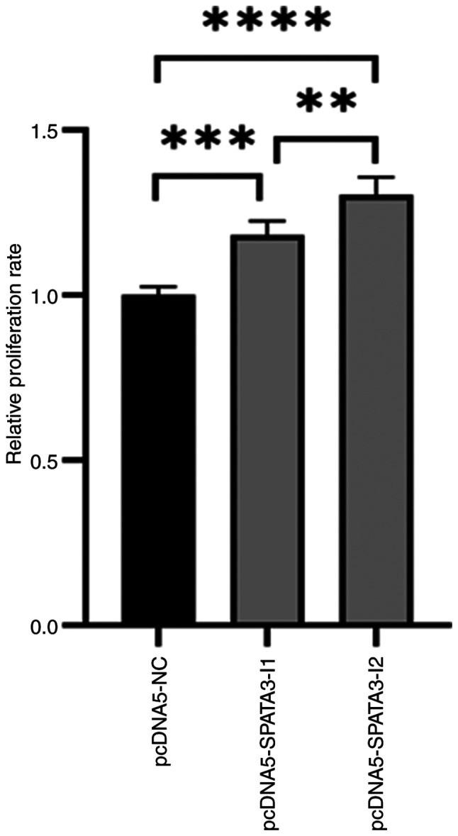
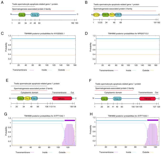
Male infertility predominantly manifests as abnormal spermatogenesis and maturation, abnormal semen quality, chromosomal abnormalities, and endocrine dysfunction. The present study aimed to explore the role, expression pattern and localization of spermatogenesis‑associated protein 3 (SPATA3) in the testis, and to analyze its spatiotemporal expression and function. Using previously acquired plasmids as templates, recombinant plasmids of different SPATA3 isoforms were constructed. Cell Counting Kit‑8 assay was used to analyze cell proliferation. Reverse transcription‑PCR and western blotting were used to determine the mRNA and protein expression levels, respectively. Different SPATA3 isoforms were induced to be overexpressed, among which SPATA3‑I1 and SPATA3‑I2 promoted cell proliferation. The subcellular localization results indicated that the green fluorescent protein fusion proteins of various isoforms were mainly localized in the nucleus. However, the fluorescence of the fusion proteins pEGFP‑C3‑SPATA3‑I3 and pEGFP‑C3‑SPATA3‑I4 tended to be distributed in the cytoplasm, accompanied by a decrease in nuclear fluorescence. Additionally, the SPATA3‑I2 isoform protein displayed notable tissue‑specific expression in testes. Notably, the SPATA3‑I2 isoform protein was not expressed in embryos or during the early development stage of the mice, yet it was highly expressed in the testes of mice aged 23‑57 days (3‑8 weeks). Immunohistochemistry revealed that the SPATA3‑I2 isoform protein was located mainly in round and elongated spermatids within the spermatogenic epithelial cells. In conclusion, the findings highlighted that SPATA3 isoforms had differential subcellular localizations and that SPATA3‑I2 exhibited a specific spatiotemporal expression pattern, suggesting its association with spermatogenesis and sperm maturation.
期刊介绍:
Molecular Medicine Reports is a monthly, peer-reviewed journal available in print and online, that includes studies devoted to molecular medicine, underscoring aspects including pharmacology, pathology, genetics, neurosciences, infectious diseases, molecular cardiology and molecular surgery. In vitro and in vivo studies of experimental model systems pertaining to the mechanisms of a variety of diseases offer researchers the necessary tools and knowledge with which to aid the diagnosis and treatment of human diseases.

 求助内容:
求助内容: 应助结果提醒方式:


