Andrea Gaggino, Leandro Inferrera, Serena Milan, Marianna Presotto, Daniele Tognetto
{"title":"伴脉络膜痣的眼息肉样脉络膜血管病变6例。","authors":"Andrea Gaggino, Leandro Inferrera, Serena Milan, Marianna Presotto, Daniele Tognetto","doi":"10.1159/000543643","DOIUrl":null,"url":null,"abstract":"<p><strong>Introduction: </strong>The aim of this study was to report the cases of 6 patients with the coexistence of polypoidal choroidal vasculopathy (PCV) and choroidal nevi.</p><p><strong>Case presentation: </strong>Six patients with the coexistence of PCV and choroidal nevi were thoroughly evaluated by slit-lamp biomicroscopy examination, color fundus photography, optical coherence tomography (OCT), OCT angiography (OCT-A), fluorescein angiography (FA), indocyanine green angiography (ICG-A), fundus blue autofluorescence (BAF), and ocular ultrasound (OU). The typical features of PCV and nevi were present in all patients, three of whom were treated with intravitreal anti-angiogenic agents. In each clinical case, the choroidal Haller's vessels adjacent to the nevus were visibly more dilated compared to normal. Color fundus photography, OCT, OCT-A, FA, ICG-A, BAF, and OU revealed similar findings across all cases. Observations revealed that choroidal nevi could instigate modifications in the outer retina, resulting in persistent alterations capable of triggering the formation of neovascularization.</p><p><strong>Conclusion: </strong>The occurrence of a PCV alongside nevus is an uncommon complication. Findings from all exams performed were consistent across all cases, highlighting the potential link between PCV and nevi.</p>","PeriodicalId":9635,"journal":{"name":"Case Reports in Ophthalmology","volume":"16 1","pages":"182-193"},"PeriodicalIF":0.6000,"publicationDate":"2025-02-19","publicationTypes":"Journal Article","fieldsOfStudy":null,"isOpenAccess":false,"openAccessPdf":"https://www.ncbi.nlm.nih.gov/pmc/articles/PMC12245153/pdf/","citationCount":"0","resultStr":"{\"title\":\"Six Cases of Polypoidal Choroidal Vasculopathy in Eyes with Choroidal Nevi.\",\"authors\":\"Andrea Gaggino, Leandro Inferrera, Serena Milan, Marianna Presotto, Daniele Tognetto\",\"doi\":\"10.1159/000543643\",\"DOIUrl\":null,\"url\":null,\"abstract\":\"<p><strong>Introduction: </strong>The aim of this study was to report the cases of 6 patients with the coexistence of polypoidal choroidal vasculopathy (PCV) and choroidal nevi.</p><p><strong>Case presentation: </strong>Six patients with the coexistence of PCV and choroidal nevi were thoroughly evaluated by slit-lamp biomicroscopy examination, color fundus photography, optical coherence tomography (OCT), OCT angiography (OCT-A), fluorescein angiography (FA), indocyanine green angiography (ICG-A), fundus blue autofluorescence (BAF), and ocular ultrasound (OU). The typical features of PCV and nevi were present in all patients, three of whom were treated with intravitreal anti-angiogenic agents. In each clinical case, the choroidal Haller's vessels adjacent to the nevus were visibly more dilated compared to normal. Color fundus photography, OCT, OCT-A, FA, ICG-A, BAF, and OU revealed similar findings across all cases. Observations revealed that choroidal nevi could instigate modifications in the outer retina, resulting in persistent alterations capable of triggering the formation of neovascularization.</p><p><strong>Conclusion: </strong>The occurrence of a PCV alongside nevus is an uncommon complication. Findings from all exams performed were consistent across all cases, highlighting the potential link between PCV and nevi.</p>\",\"PeriodicalId\":9635,\"journal\":{\"name\":\"Case Reports in Ophthalmology\",\"volume\":\"16 1\",\"pages\":\"182-193\"},\"PeriodicalIF\":0.6000,\"publicationDate\":\"2025-02-19\",\"publicationTypes\":\"Journal Article\",\"fieldsOfStudy\":null,\"isOpenAccess\":false,\"openAccessPdf\":\"https://www.ncbi.nlm.nih.gov/pmc/articles/PMC12245153/pdf/\",\"citationCount\":\"0\",\"resultStr\":null,\"platform\":\"Semanticscholar\",\"paperid\":null,\"PeriodicalName\":\"Case Reports in Ophthalmology\",\"FirstCategoryId\":\"1085\",\"ListUrlMain\":\"https://doi.org/10.1159/000543643\",\"RegionNum\":0,\"RegionCategory\":null,\"ArticlePicture\":[],\"TitleCN\":null,\"AbstractTextCN\":null,\"PMCID\":null,\"EPubDate\":\"2025/1/1 0:00:00\",\"PubModel\":\"eCollection\",\"JCR\":\"Q4\",\"JCRName\":\"OPHTHALMOLOGY\",\"Score\":null,\"Total\":0}","platform":"Semanticscholar","paperid":null,"PeriodicalName":"Case Reports in Ophthalmology","FirstCategoryId":"1085","ListUrlMain":"https://doi.org/10.1159/000543643","RegionNum":0,"RegionCategory":null,"ArticlePicture":[],"TitleCN":null,"AbstractTextCN":null,"PMCID":null,"EPubDate":"2025/1/1 0:00:00","PubModel":"eCollection","JCR":"Q4","JCRName":"OPHTHALMOLOGY","Score":null,"Total":0}
Six Cases of Polypoidal Choroidal Vasculopathy in Eyes with Choroidal Nevi.
Introduction: The aim of this study was to report the cases of 6 patients with the coexistence of polypoidal choroidal vasculopathy (PCV) and choroidal nevi.
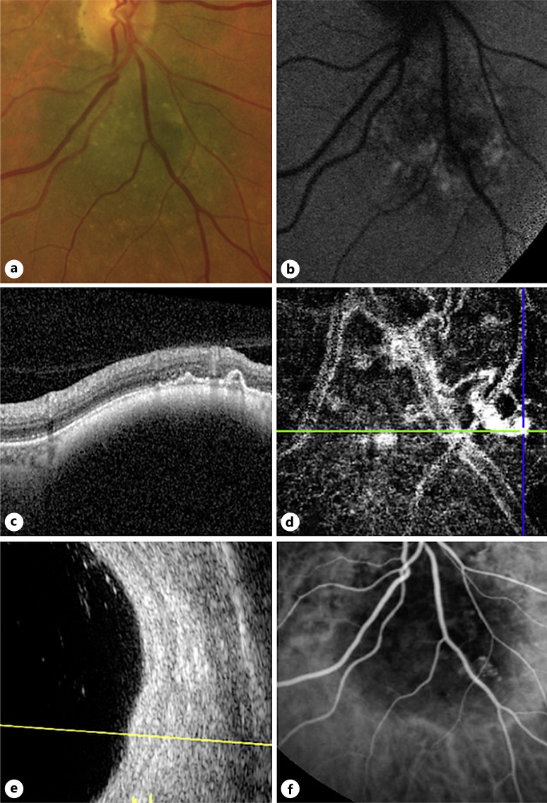
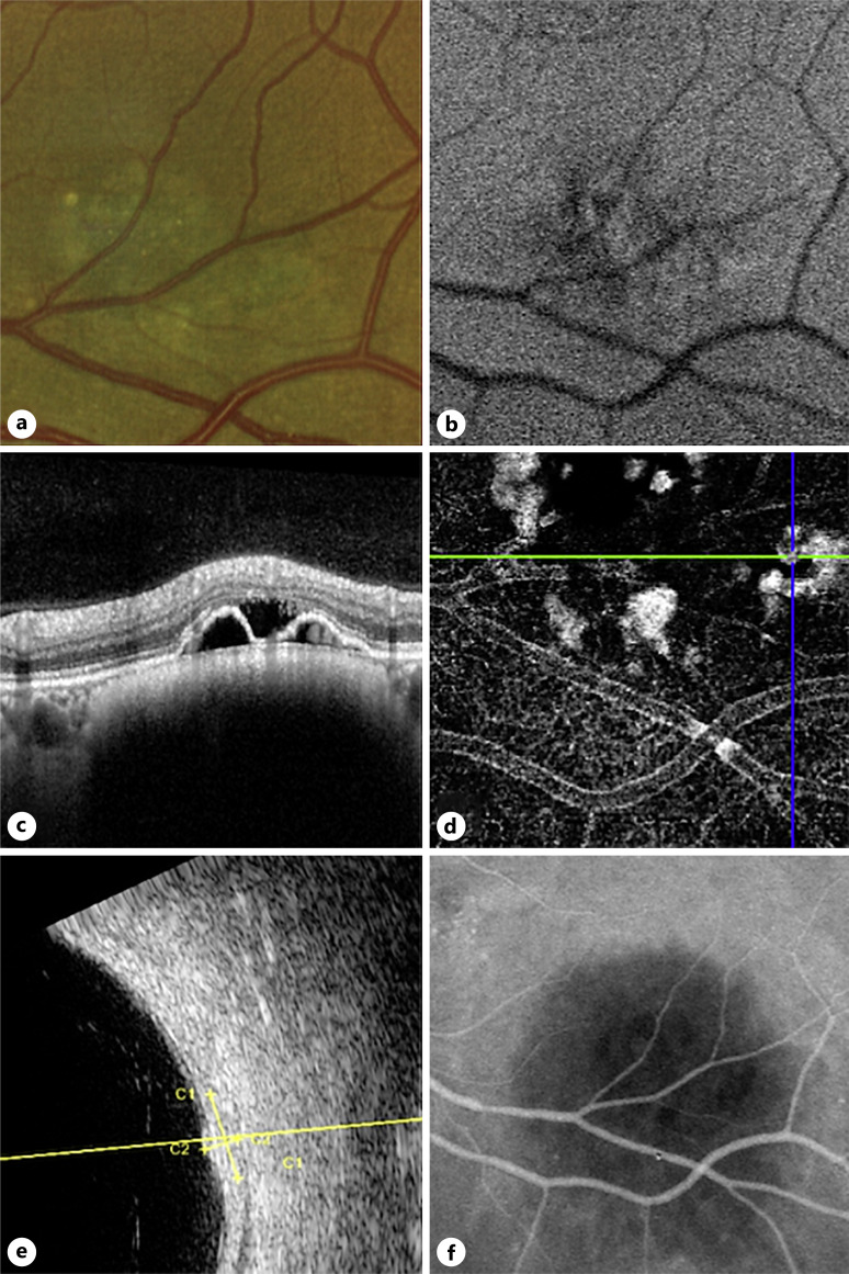
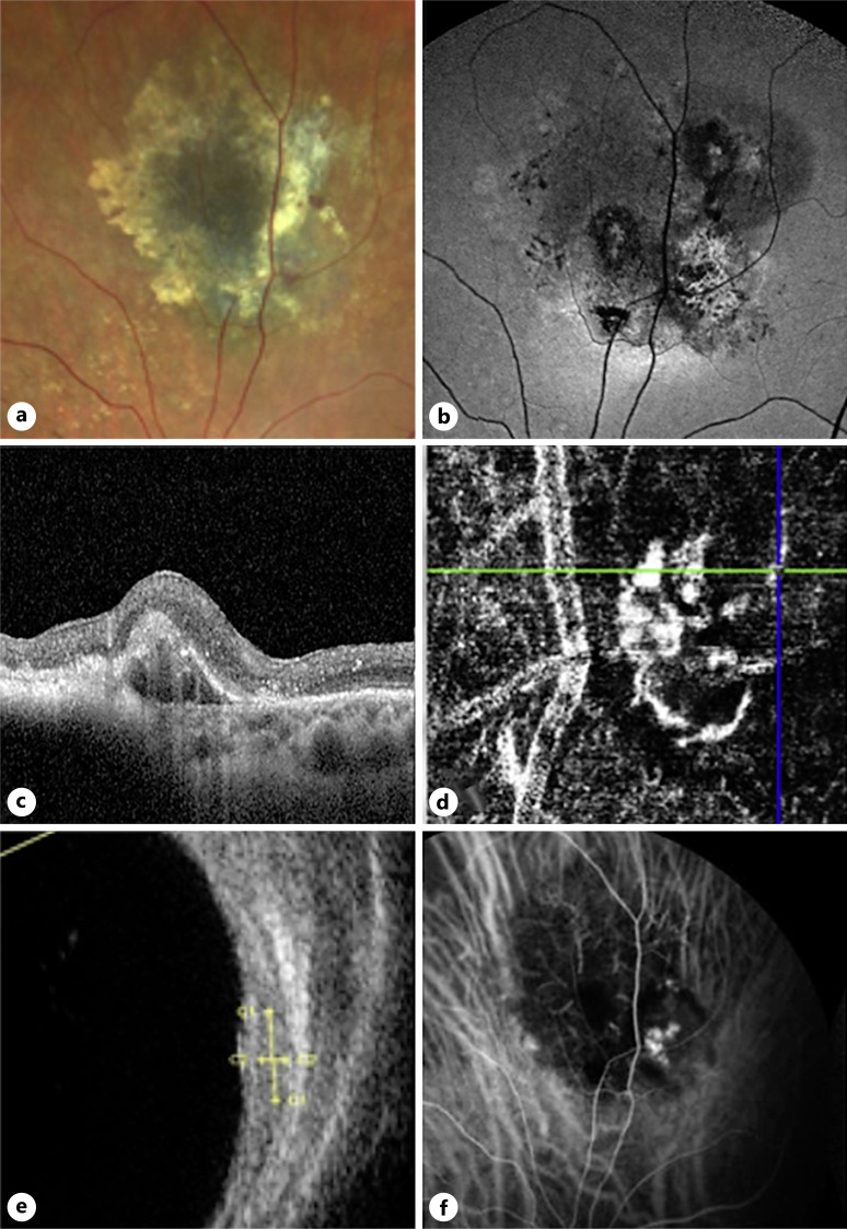
Case presentation: Six patients with the coexistence of PCV and choroidal nevi were thoroughly evaluated by slit-lamp biomicroscopy examination, color fundus photography, optical coherence tomography (OCT), OCT angiography (OCT-A), fluorescein angiography (FA), indocyanine green angiography (ICG-A), fundus blue autofluorescence (BAF), and ocular ultrasound (OU). The typical features of PCV and nevi were present in all patients, three of whom were treated with intravitreal anti-angiogenic agents. In each clinical case, the choroidal Haller's vessels adjacent to the nevus were visibly more dilated compared to normal. Color fundus photography, OCT, OCT-A, FA, ICG-A, BAF, and OU revealed similar findings across all cases. Observations revealed that choroidal nevi could instigate modifications in the outer retina, resulting in persistent alterations capable of triggering the formation of neovascularization.
Conclusion: The occurrence of a PCV alongside nevus is an uncommon complication. Findings from all exams performed were consistent across all cases, highlighting the potential link between PCV and nevi.
期刊介绍:
This peer-reviewed online-only journal publishes original case reports covering the entire spectrum of ophthalmology, including prevention, diagnosis, treatment, toxicities of therapy, supportive care, quality-of-life, and survivorship issues. The submission of negative results is strongly encouraged. The journal will also accept case reports dealing with the use of novel technologies, both in the arena of diagnosis and treatment. Supplementary material is welcomed. The intent of the journal is to provide clinicians and researchers with a tool to disseminate their personal experiences to a wider public as well as to review interesting cases encountered by colleagues all over the world. Universally used terms can be searched across the entire growing collection of case reports, further facilitating the retrieval of specific information. Following the open access principle, the entire contents can be retrieved at no charge, guaranteeing easy access to this valuable source of anecdotal information at all times.

 求助内容:
求助内容: 应助结果提醒方式:


