Stefan Stoldt, Frederike Maass, Michael Weber, Sven Dennerlein, Peter Ilgen, Jutta Gärtner, Aysenur Canfes, Sarah V. Schweighofer, Daniel C. Jans, Peter Rehling, Stefan Jakobs
{"title":"线粒体mrna的超分辨率显微镜","authors":"Stefan Stoldt, Frederike Maass, Michael Weber, Sven Dennerlein, Peter Ilgen, Jutta Gärtner, Aysenur Canfes, Sarah V. Schweighofer, Daniel C. Jans, Peter Rehling, Stefan Jakobs","doi":"10.1038/s41467-025-61577-5","DOIUrl":null,"url":null,"abstract":"<p>Mitochondria contain their own DNA (mtDNA) and a dedicated gene expression machinery. As the mitochondrial dimensions are close to the diffraction limit of classical light microscopy, the spatial distribution of mitochondrial proteins and in particular of mitochondrial mRNAs remains underexplored. Here, we establish single-molecule fluorescence in situ hybridization (smFISH) combined with STED and MINFLUX super-resolution microscopy (nanoscopy) to visualize individual mitochondrial mRNA molecules and associated proteins. STED nanoscopy reveals the spatial relationships between distinct mRNA species and proteins such as the RNA granule marker GRSF1, demonstrating adaptive changes in mRNA distribution and quantity in challenged mammalian cells and patient-derived cell lines. Notably, STED-smFISH shows the release of mRNAs during apoptosis, while MINFLUX reveals the folding of the mRNAs into variable shapes, as well as their spatial proximity to mitochondrial ribosomes. These protocols are transferable to various cell types and open new avenues for understanding mitochondrial gene regulation in health and disease.</p>","PeriodicalId":19066,"journal":{"name":"Nature Communications","volume":"22 1","pages":""},"PeriodicalIF":15.7000,"publicationDate":"2025-07-10","publicationTypes":"Journal Article","fieldsOfStudy":null,"isOpenAccess":false,"openAccessPdf":"","citationCount":"0","resultStr":"{\"title\":\"Super-resolution microscopy of mitochondrial mRNAs\",\"authors\":\"Stefan Stoldt, Frederike Maass, Michael Weber, Sven Dennerlein, Peter Ilgen, Jutta Gärtner, Aysenur Canfes, Sarah V. Schweighofer, Daniel C. Jans, Peter Rehling, Stefan Jakobs\",\"doi\":\"10.1038/s41467-025-61577-5\",\"DOIUrl\":null,\"url\":null,\"abstract\":\"<p>Mitochondria contain their own DNA (mtDNA) and a dedicated gene expression machinery. As the mitochondrial dimensions are close to the diffraction limit of classical light microscopy, the spatial distribution of mitochondrial proteins and in particular of mitochondrial mRNAs remains underexplored. Here, we establish single-molecule fluorescence in situ hybridization (smFISH) combined with STED and MINFLUX super-resolution microscopy (nanoscopy) to visualize individual mitochondrial mRNA molecules and associated proteins. STED nanoscopy reveals the spatial relationships between distinct mRNA species and proteins such as the RNA granule marker GRSF1, demonstrating adaptive changes in mRNA distribution and quantity in challenged mammalian cells and patient-derived cell lines. Notably, STED-smFISH shows the release of mRNAs during apoptosis, while MINFLUX reveals the folding of the mRNAs into variable shapes, as well as their spatial proximity to mitochondrial ribosomes. These protocols are transferable to various cell types and open new avenues for understanding mitochondrial gene regulation in health and disease.</p>\",\"PeriodicalId\":19066,\"journal\":{\"name\":\"Nature Communications\",\"volume\":\"22 1\",\"pages\":\"\"},\"PeriodicalIF\":15.7000,\"publicationDate\":\"2025-07-10\",\"publicationTypes\":\"Journal Article\",\"fieldsOfStudy\":null,\"isOpenAccess\":false,\"openAccessPdf\":\"\",\"citationCount\":\"0\",\"resultStr\":null,\"platform\":\"Semanticscholar\",\"paperid\":null,\"PeriodicalName\":\"Nature Communications\",\"FirstCategoryId\":\"103\",\"ListUrlMain\":\"https://doi.org/10.1038/s41467-025-61577-5\",\"RegionNum\":1,\"RegionCategory\":\"综合性期刊\",\"ArticlePicture\":[],\"TitleCN\":null,\"AbstractTextCN\":null,\"PMCID\":null,\"EPubDate\":\"\",\"PubModel\":\"\",\"JCR\":\"Q1\",\"JCRName\":\"MULTIDISCIPLINARY SCIENCES\",\"Score\":null,\"Total\":0}","platform":"Semanticscholar","paperid":null,"PeriodicalName":"Nature Communications","FirstCategoryId":"103","ListUrlMain":"https://doi.org/10.1038/s41467-025-61577-5","RegionNum":1,"RegionCategory":"综合性期刊","ArticlePicture":[],"TitleCN":null,"AbstractTextCN":null,"PMCID":null,"EPubDate":"","PubModel":"","JCR":"Q1","JCRName":"MULTIDISCIPLINARY SCIENCES","Score":null,"Total":0}
Super-resolution microscopy of mitochondrial mRNAs
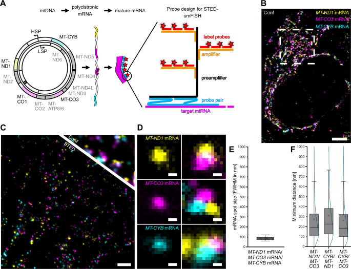
Mitochondria contain their own DNA (mtDNA) and a dedicated gene expression machinery. As the mitochondrial dimensions are close to the diffraction limit of classical light microscopy, the spatial distribution of mitochondrial proteins and in particular of mitochondrial mRNAs remains underexplored. Here, we establish single-molecule fluorescence in situ hybridization (smFISH) combined with STED and MINFLUX super-resolution microscopy (nanoscopy) to visualize individual mitochondrial mRNA molecules and associated proteins. STED nanoscopy reveals the spatial relationships between distinct mRNA species and proteins such as the RNA granule marker GRSF1, demonstrating adaptive changes in mRNA distribution and quantity in challenged mammalian cells and patient-derived cell lines. Notably, STED-smFISH shows the release of mRNAs during apoptosis, while MINFLUX reveals the folding of the mRNAs into variable shapes, as well as their spatial proximity to mitochondrial ribosomes. These protocols are transferable to various cell types and open new avenues for understanding mitochondrial gene regulation in health and disease.
期刊介绍:
Nature Communications, an open-access journal, publishes high-quality research spanning all areas of the natural sciences. Papers featured in the journal showcase significant advances relevant to specialists in each respective field. With a 2-year impact factor of 16.6 (2022) and a median time of 8 days from submission to the first editorial decision, Nature Communications is committed to rapid dissemination of research findings. As a multidisciplinary journal, it welcomes contributions from biological, health, physical, chemical, Earth, social, mathematical, applied, and engineering sciences, aiming to highlight important breakthroughs within each domain.
 求助内容:
求助内容: 应助结果提醒方式:


