Hee Sang You, Jae Yong Park, Hochan Seo, Beom Jin Kim, Jae Gyu Kim
{"title":"胃癌发生过程中液体活检样本的不同微生物特征和细胞外囊泡分析的见解。","authors":"Hee Sang You, Jae Yong Park, Hochan Seo, Beom Jin Kim, Jae Gyu Kim","doi":"10.3904/kjim.2024.339","DOIUrl":null,"url":null,"abstract":"<p><strong>Background/aims: </strong>The early detection of gastric cancer is crucial for improving patient outcomes. However, its pathogenesis is not fully understood. The microbiome and extracellular vesicles (EVs) might play a role in gastric carcinogenesis. We aimed to identify gastric-carcinogenesis-associated microbial signatures and evaluate whether these features vary across disease stages.</p><p><strong>Methods: </strong>We enrolled 141 participants (132 patients with gastric cancer or dysplasia and 9 healthy controls). Microbial-derived EVs were isolated from gastric juice, saliva, serum, and urine. Next-generation sequencing of EV-derived bacterial DNA was performed.</p><p><strong>Results: </strong>This sequencing revealed the alpha and beta diversities and microbial composition across different disease stages. The alpha diversity was significantly increased in the gastric juice and serum of disease groups. The beta diversity showed significant differences among patient groups. Distinct microbial signatures were observed across different disease stages in all four sample types. Specific bacterial species--Cutibacterium acnes, Streptococcus oralis, Pseudomonas antarctica, Ralstonia insidiosa, and Pseudomonas yamanorum--exhibited unique abundance patterns associated with disease progression, suggesting their potential as noninvasive biomarkers.</p><p><strong>Conclusion: </strong>Changes in microbial diversity and distinct microbial signatures were observed during gastric carcinogenesis in both gastric juice and extragastric samples, indicating the potential of microbial-derived EVs from liquid biopsy samples as biomarkers for gastric cancer.</p>","PeriodicalId":48785,"journal":{"name":"Korean Journal of Internal Medicine","volume":"40 4","pages":"571-583"},"PeriodicalIF":2.4000,"publicationDate":"2025-07-01","publicationTypes":"Journal Article","fieldsOfStudy":null,"isOpenAccess":false,"openAccessPdf":"https://www.ncbi.nlm.nih.gov/pmc/articles/PMC12257012/pdf/","citationCount":"0","resultStr":"{\"title\":\"Distinct microbial signatures of liquid biopsy samples during gastric carcinogenesis and insights from extracellular vesicle analysis.\",\"authors\":\"Hee Sang You, Jae Yong Park, Hochan Seo, Beom Jin Kim, Jae Gyu Kim\",\"doi\":\"10.3904/kjim.2024.339\",\"DOIUrl\":null,\"url\":null,\"abstract\":\"<p><strong>Background/aims: </strong>The early detection of gastric cancer is crucial for improving patient outcomes. However, its pathogenesis is not fully understood. The microbiome and extracellular vesicles (EVs) might play a role in gastric carcinogenesis. We aimed to identify gastric-carcinogenesis-associated microbial signatures and evaluate whether these features vary across disease stages.</p><p><strong>Methods: </strong>We enrolled 141 participants (132 patients with gastric cancer or dysplasia and 9 healthy controls). Microbial-derived EVs were isolated from gastric juice, saliva, serum, and urine. Next-generation sequencing of EV-derived bacterial DNA was performed.</p><p><strong>Results: </strong>This sequencing revealed the alpha and beta diversities and microbial composition across different disease stages. The alpha diversity was significantly increased in the gastric juice and serum of disease groups. The beta diversity showed significant differences among patient groups. Distinct microbial signatures were observed across different disease stages in all four sample types. Specific bacterial species--Cutibacterium acnes, Streptococcus oralis, Pseudomonas antarctica, Ralstonia insidiosa, and Pseudomonas yamanorum--exhibited unique abundance patterns associated with disease progression, suggesting their potential as noninvasive biomarkers.</p><p><strong>Conclusion: </strong>Changes in microbial diversity and distinct microbial signatures were observed during gastric carcinogenesis in both gastric juice and extragastric samples, indicating the potential of microbial-derived EVs from liquid biopsy samples as biomarkers for gastric cancer.</p>\",\"PeriodicalId\":48785,\"journal\":{\"name\":\"Korean Journal of Internal Medicine\",\"volume\":\"40 4\",\"pages\":\"571-583\"},\"PeriodicalIF\":2.4000,\"publicationDate\":\"2025-07-01\",\"publicationTypes\":\"Journal Article\",\"fieldsOfStudy\":null,\"isOpenAccess\":false,\"openAccessPdf\":\"https://www.ncbi.nlm.nih.gov/pmc/articles/PMC12257012/pdf/\",\"citationCount\":\"0\",\"resultStr\":null,\"platform\":\"Semanticscholar\",\"paperid\":null,\"PeriodicalName\":\"Korean Journal of Internal Medicine\",\"FirstCategoryId\":\"3\",\"ListUrlMain\":\"https://doi.org/10.3904/kjim.2024.339\",\"RegionNum\":4,\"RegionCategory\":\"医学\",\"ArticlePicture\":[],\"TitleCN\":null,\"AbstractTextCN\":null,\"PMCID\":null,\"EPubDate\":\"\",\"PubModel\":\"\",\"JCR\":\"Q2\",\"JCRName\":\"MEDICINE, GENERAL & INTERNAL\",\"Score\":null,\"Total\":0}","platform":"Semanticscholar","paperid":null,"PeriodicalName":"Korean Journal of Internal Medicine","FirstCategoryId":"3","ListUrlMain":"https://doi.org/10.3904/kjim.2024.339","RegionNum":4,"RegionCategory":"医学","ArticlePicture":[],"TitleCN":null,"AbstractTextCN":null,"PMCID":null,"EPubDate":"","PubModel":"","JCR":"Q2","JCRName":"MEDICINE, GENERAL & INTERNAL","Score":null,"Total":0}
Distinct microbial signatures of liquid biopsy samples during gastric carcinogenesis and insights from extracellular vesicle analysis.
Background/aims: The early detection of gastric cancer is crucial for improving patient outcomes. However, its pathogenesis is not fully understood. The microbiome and extracellular vesicles (EVs) might play a role in gastric carcinogenesis. We aimed to identify gastric-carcinogenesis-associated microbial signatures and evaluate whether these features vary across disease stages.
Methods: We enrolled 141 participants (132 patients with gastric cancer or dysplasia and 9 healthy controls). Microbial-derived EVs were isolated from gastric juice, saliva, serum, and urine. Next-generation sequencing of EV-derived bacterial DNA was performed.
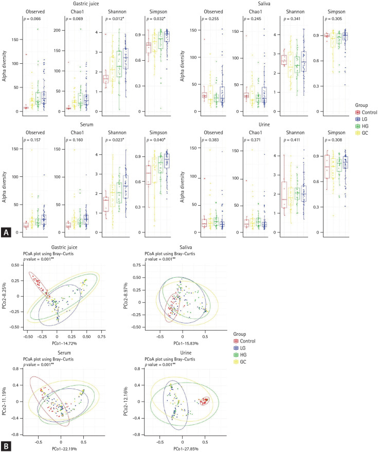
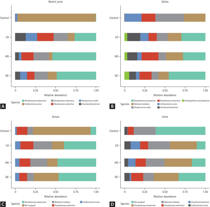
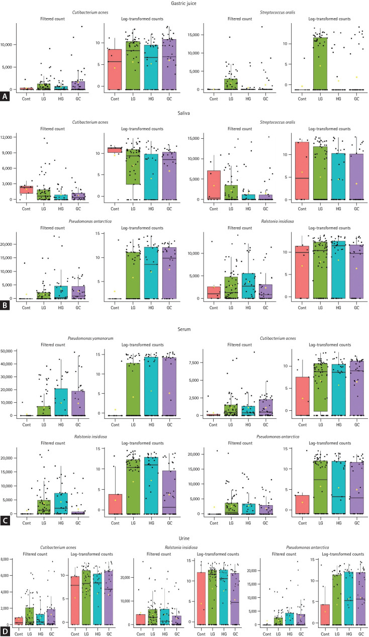
Results: This sequencing revealed the alpha and beta diversities and microbial composition across different disease stages. The alpha diversity was significantly increased in the gastric juice and serum of disease groups. The beta diversity showed significant differences among patient groups. Distinct microbial signatures were observed across different disease stages in all four sample types. Specific bacterial species--Cutibacterium acnes, Streptococcus oralis, Pseudomonas antarctica, Ralstonia insidiosa, and Pseudomonas yamanorum--exhibited unique abundance patterns associated with disease progression, suggesting their potential as noninvasive biomarkers.
Conclusion: Changes in microbial diversity and distinct microbial signatures were observed during gastric carcinogenesis in both gastric juice and extragastric samples, indicating the potential of microbial-derived EVs from liquid biopsy samples as biomarkers for gastric cancer.
期刊介绍:
The Korean Journal of Internal Medicine is an international medical journal published in English by the Korean Association of Internal Medicine. The Journal publishes peer-reviewed original articles, reviews, and editorials on all aspects of medicine, including clinical investigations and basic research. Both human and experimental animal studies are welcome, as are new findings on the epidemiology, pathogenesis, diagnosis, and treatment of diseases. Case reports will be published only in exceptional circumstances, when they illustrate a rare occurrence of clinical importance. Letters to the editor are encouraged for specific comments on published articles and general viewpoints.
 求助内容:
求助内容: 应助结果提醒方式:


