{"title":"膀胱过度活动大鼠脊神经刺激时脑活动和功能连通性的变化。","authors":"Haoyu Sun, Yongheng Zhou, Qinggang Liu, Xing Li, Limin Liao","doi":"10.5213/inj.2448420.210","DOIUrl":null,"url":null,"abstract":"<p><strong>Purpose: </strong>Sacral neuromodulation is widely used for refractory overactive bladder; however, its mechanism of action remains unclear. This study aims to investigate real-time changes in brain activity and functional connectivity (FC) during neuromodulation in an overactive bladder (OAB) rat model using functional magnetic resonance imaging.</p><p><strong>Methods: </strong>Twelve female Sprague Dawley rats were implanted with fine bipolar electrodes adjacent to the L6 nerve root. Cystometry was performed on normal rats, acetic acid-induced OAB rats, and during spinal nerve stimulation (SNS) to confirm efficacy. Task-based functional magnetic resonance imaging (fMRI) was acquired using a 20-second rest-stimulus cycle, followed by T2-weighted anatomical imaging on a 9.4T MRI scanner. Comparative analyses examined changes in the amplitude of low-frequency fluctuations (ALFF) and FC between normal and OAB rats. Brain activity during SNS was further assessed using the general linear model (GLM) and FC analysis. Statistical significance was defined as P<0.05 (family-wise error-corrected).</p><p><strong>Results: </strong>Compared with normal rats, OAB rats exhibited increased ALFF in the left prefrontal cortex, periaqueductal gray (PAG), and left primary somatosensory cortex. In addition, FC between the PAG and pons was enhanced (P=0.002). GLM analysis revealed that the left primary somatosensory cortex, left prefrontal cortex, corpus callosum, left secondary motor area, and right brainstem exhibited decreased activity during SNS (P<0.05). Significant FC changes were observed between several regions: the left cerebellum and left caudal zona incerta (P=0.024), right fasciculus retroflexus and left ventral orbital area (P=0.025), and between the pons and PAG (P=0.004). Seed-to-voxel analysis indicated altered FC between clusters identified in the GLM analysis and regions including the PAG, left cingulate area, left prefrontal cortex, left caudate putamen, and right granular insular cortex.</p><p><strong>Conclusion: </strong>Our fMRI study identified several alterations in brain activity during SNS in rats. Specifically, activity in the left prefrontal cortex decreased during SNS, and FC between the PAG and pons was reduced. These changes may represent central mechanisms underlying sacral neuromodulation in OAB patients.</p>","PeriodicalId":14466,"journal":{"name":"International Neurourology Journal","volume":"29 2","pages":"81-91"},"PeriodicalIF":2.1000,"publicationDate":"2025-06-01","publicationTypes":"Journal Article","fieldsOfStudy":null,"isOpenAccess":false,"openAccessPdf":"https://www.ncbi.nlm.nih.gov/pmc/articles/PMC12242188/pdf/","citationCount":"0","resultStr":"{\"title\":\"Changes in Brain Activity and Functional Connectivity During Spinal Nerve Stimulation in a Rat Model of Overactive Bladder.\",\"authors\":\"Haoyu Sun, Yongheng Zhou, Qinggang Liu, Xing Li, Limin Liao\",\"doi\":\"10.5213/inj.2448420.210\",\"DOIUrl\":null,\"url\":null,\"abstract\":\"<p><strong>Purpose: </strong>Sacral neuromodulation is widely used for refractory overactive bladder; however, its mechanism of action remains unclear. This study aims to investigate real-time changes in brain activity and functional connectivity (FC) during neuromodulation in an overactive bladder (OAB) rat model using functional magnetic resonance imaging.</p><p><strong>Methods: </strong>Twelve female Sprague Dawley rats were implanted with fine bipolar electrodes adjacent to the L6 nerve root. Cystometry was performed on normal rats, acetic acid-induced OAB rats, and during spinal nerve stimulation (SNS) to confirm efficacy. Task-based functional magnetic resonance imaging (fMRI) was acquired using a 20-second rest-stimulus cycle, followed by T2-weighted anatomical imaging on a 9.4T MRI scanner. Comparative analyses examined changes in the amplitude of low-frequency fluctuations (ALFF) and FC between normal and OAB rats. Brain activity during SNS was further assessed using the general linear model (GLM) and FC analysis. Statistical significance was defined as P<0.05 (family-wise error-corrected).</p><p><strong>Results: </strong>Compared with normal rats, OAB rats exhibited increased ALFF in the left prefrontal cortex, periaqueductal gray (PAG), and left primary somatosensory cortex. In addition, FC between the PAG and pons was enhanced (P=0.002). GLM analysis revealed that the left primary somatosensory cortex, left prefrontal cortex, corpus callosum, left secondary motor area, and right brainstem exhibited decreased activity during SNS (P<0.05). Significant FC changes were observed between several regions: the left cerebellum and left caudal zona incerta (P=0.024), right fasciculus retroflexus and left ventral orbital area (P=0.025), and between the pons and PAG (P=0.004). Seed-to-voxel analysis indicated altered FC between clusters identified in the GLM analysis and regions including the PAG, left cingulate area, left prefrontal cortex, left caudate putamen, and right granular insular cortex.</p><p><strong>Conclusion: </strong>Our fMRI study identified several alterations in brain activity during SNS in rats. Specifically, activity in the left prefrontal cortex decreased during SNS, and FC between the PAG and pons was reduced. These changes may represent central mechanisms underlying sacral neuromodulation in OAB patients.</p>\",\"PeriodicalId\":14466,\"journal\":{\"name\":\"International Neurourology Journal\",\"volume\":\"29 2\",\"pages\":\"81-91\"},\"PeriodicalIF\":2.1000,\"publicationDate\":\"2025-06-01\",\"publicationTypes\":\"Journal Article\",\"fieldsOfStudy\":null,\"isOpenAccess\":false,\"openAccessPdf\":\"https://www.ncbi.nlm.nih.gov/pmc/articles/PMC12242188/pdf/\",\"citationCount\":\"0\",\"resultStr\":null,\"platform\":\"Semanticscholar\",\"paperid\":null,\"PeriodicalName\":\"International Neurourology Journal\",\"FirstCategoryId\":\"3\",\"ListUrlMain\":\"https://doi.org/10.5213/inj.2448420.210\",\"RegionNum\":3,\"RegionCategory\":\"医学\",\"ArticlePicture\":[],\"TitleCN\":null,\"AbstractTextCN\":null,\"PMCID\":null,\"EPubDate\":\"2025/6/30 0:00:00\",\"PubModel\":\"Epub\",\"JCR\":\"Q3\",\"JCRName\":\"UROLOGY & NEPHROLOGY\",\"Score\":null,\"Total\":0}","platform":"Semanticscholar","paperid":null,"PeriodicalName":"International Neurourology Journal","FirstCategoryId":"3","ListUrlMain":"https://doi.org/10.5213/inj.2448420.210","RegionNum":3,"RegionCategory":"医学","ArticlePicture":[],"TitleCN":null,"AbstractTextCN":null,"PMCID":null,"EPubDate":"2025/6/30 0:00:00","PubModel":"Epub","JCR":"Q3","JCRName":"UROLOGY & NEPHROLOGY","Score":null,"Total":0}
Changes in Brain Activity and Functional Connectivity During Spinal Nerve Stimulation in a Rat Model of Overactive Bladder.
Purpose: Sacral neuromodulation is widely used for refractory overactive bladder; however, its mechanism of action remains unclear. This study aims to investigate real-time changes in brain activity and functional connectivity (FC) during neuromodulation in an overactive bladder (OAB) rat model using functional magnetic resonance imaging.
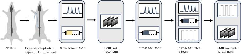
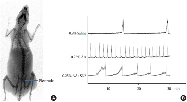
Methods: Twelve female Sprague Dawley rats were implanted with fine bipolar electrodes adjacent to the L6 nerve root. Cystometry was performed on normal rats, acetic acid-induced OAB rats, and during spinal nerve stimulation (SNS) to confirm efficacy. Task-based functional magnetic resonance imaging (fMRI) was acquired using a 20-second rest-stimulus cycle, followed by T2-weighted anatomical imaging on a 9.4T MRI scanner. Comparative analyses examined changes in the amplitude of low-frequency fluctuations (ALFF) and FC between normal and OAB rats. Brain activity during SNS was further assessed using the general linear model (GLM) and FC analysis. Statistical significance was defined as P<0.05 (family-wise error-corrected).
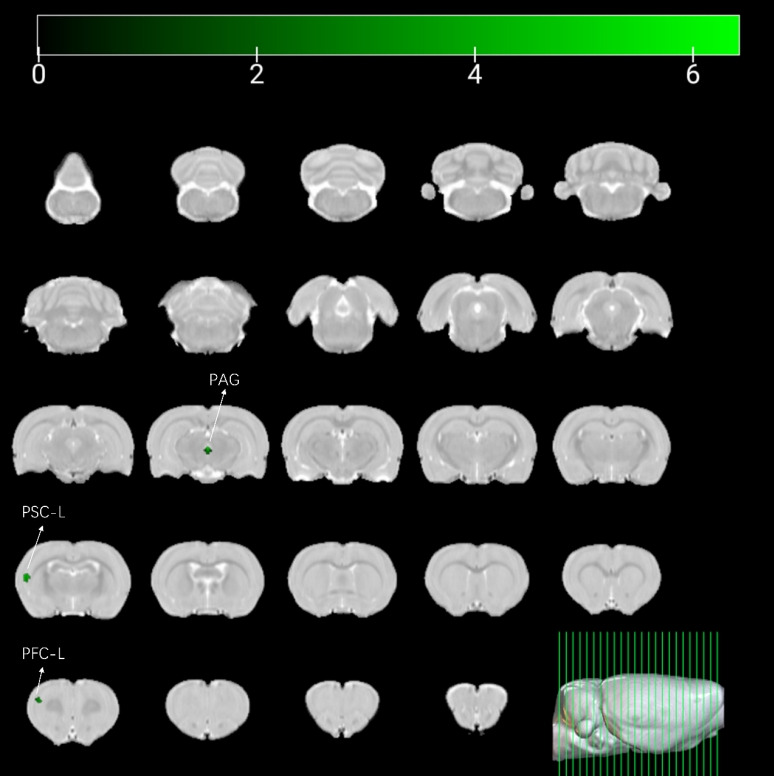
Results: Compared with normal rats, OAB rats exhibited increased ALFF in the left prefrontal cortex, periaqueductal gray (PAG), and left primary somatosensory cortex. In addition, FC between the PAG and pons was enhanced (P=0.002). GLM analysis revealed that the left primary somatosensory cortex, left prefrontal cortex, corpus callosum, left secondary motor area, and right brainstem exhibited decreased activity during SNS (P<0.05). Significant FC changes were observed between several regions: the left cerebellum and left caudal zona incerta (P=0.024), right fasciculus retroflexus and left ventral orbital area (P=0.025), and between the pons and PAG (P=0.004). Seed-to-voxel analysis indicated altered FC between clusters identified in the GLM analysis and regions including the PAG, left cingulate area, left prefrontal cortex, left caudate putamen, and right granular insular cortex.
Conclusion: Our fMRI study identified several alterations in brain activity during SNS in rats. Specifically, activity in the left prefrontal cortex decreased during SNS, and FC between the PAG and pons was reduced. These changes may represent central mechanisms underlying sacral neuromodulation in OAB patients.
期刊介绍:
The International Neurourology Journal (Int Neurourol J, INJ) is a quarterly international journal that publishes high-quality research papers that provide the most significant and promising achievements in the fields of clinical neurourology and fundamental science. Specifically, fundamental science includes the most influential research papers from all fields of science and technology, revolutionizing what physicians and researchers practicing the art of neurourology worldwide know. Thus, we welcome valuable basic research articles to introduce cutting-edge translational research of fundamental sciences to clinical neurourology. In the editorials, urologists will present their perspectives on these articles. The original mission statement of the INJ was published on October 12, 1997.
INJ provides authors a fast review of their work and makes a decision in an average of three to four weeks of receiving submissions. If accepted, articles are posted online in fully citable form. Supplementary issues will be published interim to quarterlies, as necessary, to fully allow berth to accept and publish relevant articles.
 求助内容:
求助内容: 应助结果提醒方式:


