封面图片
IF 1.6
4区 生物学
Q4 CELL BIOLOGY
引用次数: 0
摘要
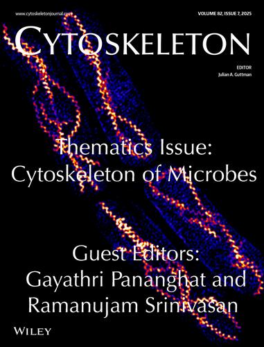
封面:图片显示幽门螺杆菌FtsZ-GFP (HpFtsZ-GFP)在分裂酵母——裂糖酵母(Schizosaccharomyces pombe)中表达成螺旋结构。采用三维结构照明显微镜(3D-SIM)对结构进行成像,得到最大强度投影图像。来源:Sakshi Poddar和Ramanujam Srinivasan(印度国家科学教育与研究所生物科学学院)。本文章由计算机程序翻译,如有差异,请以英文原文为准。
Front Cover Image
ON THE FRONT COVER: The image shows the assembly of Helicobacter pylori FtsZ-GFP (HpFtsZ-GFP) into spiral structures when expressed in fission yeast, Schizosaccharomyces pombe. Three-dimensional structured illumination microscopy (3D-SIM) was used to image the structures and maximum intensity projection images are shown.
Credit: Sakshi Poddar and Ramanujam Srinivasan (School of Biological Sciences, National Institute of Science Education and Research, India).
求助全文
通过发布文献求助,成功后即可免费获取论文全文。
去求助
来源期刊
Cytoskeleton
CELL BIOLOGY-
CiteScore
5.50
自引率
3.40%
发文量
24
审稿时长
6-12 weeks
期刊介绍:
Cytoskeleton focuses on all aspects of cytoskeletal research in healthy and diseased states, spanning genetic and cell biological observations, biochemical, biophysical and structural studies, mathematical modeling and theory. This includes, but is certainly not limited to, classic polymer systems of eukaryotic cells and their structural sites of attachment on membranes and organelles, as well as the bacterial cytoskeleton, the nucleoskeleton, and uncoventional polymer systems with structural/organizational roles. Cytoskeleton is published in 12 issues annually, and special issues will be dedicated to especially-active or newly-emerging areas of cytoskeletal research.
 求助内容:
求助内容: 应助结果提醒方式:


