Laith Hamood Aswad Al-Salmany, Lara Kusrat Hussein, Zena Hekmat Altaee
{"title":"费卢杰市正畸样本中犬类患病率、定位及相关病因分析。","authors":"Laith Hamood Aswad Al-Salmany, Lara Kusrat Hussein, Zena Hekmat Altaee","doi":"10.4103/jos.jos_109_24","DOIUrl":null,"url":null,"abstract":"<p><strong>Objective: </strong>Canine impaction isn't uncommon malocclusion and their treatment is complex and annoying to both patient and orthodontist. This study was aimed to determine the prevalence of impacted canine in orthodontic patients of Fallujah city with determining the related etiological factors.</p><p><strong>Materials and methods: </strong>The study consisted of 590 patients (412 females and 178 males) coming to the clinic for orthodontic treatment. A panoramic radiograph was taken for each subject. Cone beam computed tomography was taken for each patient with an impacted canine for accurate localization of trapped teeth. Patients with impacted canines were examined for number, distribution, crown depth, angulation and apical position of impacted teeth, lateral incisor condition, and presence of associated local factors.</p><p><strong>Results: </strong>For the total sample, forty patients (40) had impacted canines with a mean age of 22.6 years. Female to male ratio was 1.5:1. The maxilla represented 80.6% of total impaction cases. Palatally impacted canines represented 72.6% and were mostly seen bilaterally. The mandible formed 19.4% of impacted canines. Mesially and vertically angulated impacted teeth represented the majority of the cases for upper and lower jaws respectively. The impaction depth was recognized as D2 at the maxilla and D1 at the mandible as the commonest impaction level. Apical mislocation was presented in 21% of cases.</p><p><strong>Conclusion: </strong>The prevalence of impacted canines was 6.8%. Retained primary canines were associated with 76% of total impacted canines. About 32% of total maxillary impacted canines were associated with anomalous or missing lateral incisors.</p>","PeriodicalId":16604,"journal":{"name":"Journal of Orthodontic Science","volume":"14 ","pages":"15"},"PeriodicalIF":0.0000,"publicationDate":"2025-06-10","publicationTypes":"Journal Article","fieldsOfStudy":null,"isOpenAccess":false,"openAccessPdf":"https://www.ncbi.nlm.nih.gov/pmc/articles/PMC12236999/pdf/","citationCount":"0","resultStr":"{\"title\":\"Impacted canine prevalence, localization, and related etiological factors among orthodontic sample of Fallujah city.\",\"authors\":\"Laith Hamood Aswad Al-Salmany, Lara Kusrat Hussein, Zena Hekmat Altaee\",\"doi\":\"10.4103/jos.jos_109_24\",\"DOIUrl\":null,\"url\":null,\"abstract\":\"<p><strong>Objective: </strong>Canine impaction isn't uncommon malocclusion and their treatment is complex and annoying to both patient and orthodontist. This study was aimed to determine the prevalence of impacted canine in orthodontic patients of Fallujah city with determining the related etiological factors.</p><p><strong>Materials and methods: </strong>The study consisted of 590 patients (412 females and 178 males) coming to the clinic for orthodontic treatment. A panoramic radiograph was taken for each subject. Cone beam computed tomography was taken for each patient with an impacted canine for accurate localization of trapped teeth. Patients with impacted canines were examined for number, distribution, crown depth, angulation and apical position of impacted teeth, lateral incisor condition, and presence of associated local factors.</p><p><strong>Results: </strong>For the total sample, forty patients (40) had impacted canines with a mean age of 22.6 years. Female to male ratio was 1.5:1. The maxilla represented 80.6% of total impaction cases. Palatally impacted canines represented 72.6% and were mostly seen bilaterally. The mandible formed 19.4% of impacted canines. Mesially and vertically angulated impacted teeth represented the majority of the cases for upper and lower jaws respectively. The impaction depth was recognized as D2 at the maxilla and D1 at the mandible as the commonest impaction level. Apical mislocation was presented in 21% of cases.</p><p><strong>Conclusion: </strong>The prevalence of impacted canines was 6.8%. Retained primary canines were associated with 76% of total impacted canines. About 32% of total maxillary impacted canines were associated with anomalous or missing lateral incisors.</p>\",\"PeriodicalId\":16604,\"journal\":{\"name\":\"Journal of Orthodontic Science\",\"volume\":\"14 \",\"pages\":\"15\"},\"PeriodicalIF\":0.0000,\"publicationDate\":\"2025-06-10\",\"publicationTypes\":\"Journal Article\",\"fieldsOfStudy\":null,\"isOpenAccess\":false,\"openAccessPdf\":\"https://www.ncbi.nlm.nih.gov/pmc/articles/PMC12236999/pdf/\",\"citationCount\":\"0\",\"resultStr\":null,\"platform\":\"Semanticscholar\",\"paperid\":null,\"PeriodicalName\":\"Journal of Orthodontic Science\",\"FirstCategoryId\":\"1085\",\"ListUrlMain\":\"https://doi.org/10.4103/jos.jos_109_24\",\"RegionNum\":0,\"RegionCategory\":null,\"ArticlePicture\":[],\"TitleCN\":null,\"AbstractTextCN\":null,\"PMCID\":null,\"EPubDate\":\"2025/1/1 0:00:00\",\"PubModel\":\"eCollection\",\"JCR\":\"Q2\",\"JCRName\":\"Dentistry\",\"Score\":null,\"Total\":0}","platform":"Semanticscholar","paperid":null,"PeriodicalName":"Journal of Orthodontic Science","FirstCategoryId":"1085","ListUrlMain":"https://doi.org/10.4103/jos.jos_109_24","RegionNum":0,"RegionCategory":null,"ArticlePicture":[],"TitleCN":null,"AbstractTextCN":null,"PMCID":null,"EPubDate":"2025/1/1 0:00:00","PubModel":"eCollection","JCR":"Q2","JCRName":"Dentistry","Score":null,"Total":0}
Impacted canine prevalence, localization, and related etiological factors among orthodontic sample of Fallujah city.
Objective: Canine impaction isn't uncommon malocclusion and their treatment is complex and annoying to both patient and orthodontist. This study was aimed to determine the prevalence of impacted canine in orthodontic patients of Fallujah city with determining the related etiological factors.
Materials and methods: The study consisted of 590 patients (412 females and 178 males) coming to the clinic for orthodontic treatment. A panoramic radiograph was taken for each subject. Cone beam computed tomography was taken for each patient with an impacted canine for accurate localization of trapped teeth. Patients with impacted canines were examined for number, distribution, crown depth, angulation and apical position of impacted teeth, lateral incisor condition, and presence of associated local factors.
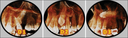
Results: For the total sample, forty patients (40) had impacted canines with a mean age of 22.6 years. Female to male ratio was 1.5:1. The maxilla represented 80.6% of total impaction cases. Palatally impacted canines represented 72.6% and were mostly seen bilaterally. The mandible formed 19.4% of impacted canines. Mesially and vertically angulated impacted teeth represented the majority of the cases for upper and lower jaws respectively. The impaction depth was recognized as D2 at the maxilla and D1 at the mandible as the commonest impaction level. Apical mislocation was presented in 21% of cases.
Conclusion: The prevalence of impacted canines was 6.8%. Retained primary canines were associated with 76% of total impacted canines. About 32% of total maxillary impacted canines were associated with anomalous or missing lateral incisors.

 求助内容:
求助内容: 应助结果提醒方式:


