Di Li, Peikang Wang, Man Zhang, Xinkai Zhang, Hailun Yao, Xing Liu
{"title":"青少年特发性脊柱侧凸检查方法研究进展。","authors":"Di Li, Peikang Wang, Man Zhang, Xinkai Zhang, Hailun Yao, Xing Liu","doi":"10.1002/pdi3.2518","DOIUrl":null,"url":null,"abstract":"<p><p>The purpose of this article is to provide an overview of techniques for evaluating patients with adolescent idiopathic scoliosis (AIS). It encompasses the history, clinical examinations, and diagnostic imaging methods for AIS. These methods include digital radiological examination, EOS® imaging, nuclear medicine, ultrasound, body surface topography techniques such as the Moiré pattern technique, raster stereophotography, and DIERS formetric 4D as well as computed tomography and magnetic resonance imaging (MRI). Traditionally, full-spine standing X-rays have been the standard for diagnosing AIS. High-quality clinical assessments may continue as a reference for assessing other spinal deformities. However, the new diagnostic imaging methods aim to reduce radiation exposure while maintaining image quality and practicality. Emerging technologies demonstrate strong reliability and effectiveness in diagnostic imaging of AlS. These techniques may be beneficial for diagnosing and monitoring AIS and its progression without requiring high levels of radiation exposure. The article is a search and summary of the PubMed electronic database to understand the current and future status of AIS imaging technology, which can not only help to introduce other researchers to the field but also serve as a valuable source for healthcare professionals to study existing methods, develop new ones, or select evaluation strategies.</p>","PeriodicalId":520221,"journal":{"name":"Pediatric discovery","volume":"3 1","pages":"e2518"},"PeriodicalIF":0.0000,"publicationDate":"2025-01-09","publicationTypes":"Journal Article","fieldsOfStudy":null,"isOpenAccess":false,"openAccessPdf":"https://www.ncbi.nlm.nih.gov/pmc/articles/PMC12118105/pdf/","citationCount":"0","resultStr":"{\"title\":\"Advances in examination methods for adolescent idiopathic scoliosis.\",\"authors\":\"Di Li, Peikang Wang, Man Zhang, Xinkai Zhang, Hailun Yao, Xing Liu\",\"doi\":\"10.1002/pdi3.2518\",\"DOIUrl\":null,\"url\":null,\"abstract\":\"<p><p>The purpose of this article is to provide an overview of techniques for evaluating patients with adolescent idiopathic scoliosis (AIS). It encompasses the history, clinical examinations, and diagnostic imaging methods for AIS. These methods include digital radiological examination, EOS® imaging, nuclear medicine, ultrasound, body surface topography techniques such as the Moiré pattern technique, raster stereophotography, and DIERS formetric 4D as well as computed tomography and magnetic resonance imaging (MRI). Traditionally, full-spine standing X-rays have been the standard for diagnosing AIS. High-quality clinical assessments may continue as a reference for assessing other spinal deformities. However, the new diagnostic imaging methods aim to reduce radiation exposure while maintaining image quality and practicality. Emerging technologies demonstrate strong reliability and effectiveness in diagnostic imaging of AlS. These techniques may be beneficial for diagnosing and monitoring AIS and its progression without requiring high levels of radiation exposure. The article is a search and summary of the PubMed electronic database to understand the current and future status of AIS imaging technology, which can not only help to introduce other researchers to the field but also serve as a valuable source for healthcare professionals to study existing methods, develop new ones, or select evaluation strategies.</p>\",\"PeriodicalId\":520221,\"journal\":{\"name\":\"Pediatric discovery\",\"volume\":\"3 1\",\"pages\":\"e2518\"},\"PeriodicalIF\":0.0000,\"publicationDate\":\"2025-01-09\",\"publicationTypes\":\"Journal Article\",\"fieldsOfStudy\":null,\"isOpenAccess\":false,\"openAccessPdf\":\"https://www.ncbi.nlm.nih.gov/pmc/articles/PMC12118105/pdf/\",\"citationCount\":\"0\",\"resultStr\":null,\"platform\":\"Semanticscholar\",\"paperid\":null,\"PeriodicalName\":\"Pediatric discovery\",\"FirstCategoryId\":\"1085\",\"ListUrlMain\":\"https://doi.org/10.1002/pdi3.2518\",\"RegionNum\":0,\"RegionCategory\":null,\"ArticlePicture\":[],\"TitleCN\":null,\"AbstractTextCN\":null,\"PMCID\":null,\"EPubDate\":\"2025/3/1 0:00:00\",\"PubModel\":\"eCollection\",\"JCR\":\"\",\"JCRName\":\"\",\"Score\":null,\"Total\":0}","platform":"Semanticscholar","paperid":null,"PeriodicalName":"Pediatric discovery","FirstCategoryId":"1085","ListUrlMain":"https://doi.org/10.1002/pdi3.2518","RegionNum":0,"RegionCategory":null,"ArticlePicture":[],"TitleCN":null,"AbstractTextCN":null,"PMCID":null,"EPubDate":"2025/3/1 0:00:00","PubModel":"eCollection","JCR":"","JCRName":"","Score":null,"Total":0}
Advances in examination methods for adolescent idiopathic scoliosis.
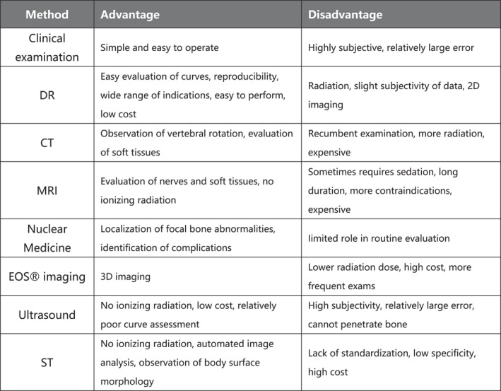
The purpose of this article is to provide an overview of techniques for evaluating patients with adolescent idiopathic scoliosis (AIS). It encompasses the history, clinical examinations, and diagnostic imaging methods for AIS. These methods include digital radiological examination, EOS® imaging, nuclear medicine, ultrasound, body surface topography techniques such as the Moiré pattern technique, raster stereophotography, and DIERS formetric 4D as well as computed tomography and magnetic resonance imaging (MRI). Traditionally, full-spine standing X-rays have been the standard for diagnosing AIS. High-quality clinical assessments may continue as a reference for assessing other spinal deformities. However, the new diagnostic imaging methods aim to reduce radiation exposure while maintaining image quality and practicality. Emerging technologies demonstrate strong reliability and effectiveness in diagnostic imaging of AlS. These techniques may be beneficial for diagnosing and monitoring AIS and its progression without requiring high levels of radiation exposure. The article is a search and summary of the PubMed electronic database to understand the current and future status of AIS imaging technology, which can not only help to introduce other researchers to the field but also serve as a valuable source for healthcare professionals to study existing methods, develop new ones, or select evaluation strategies.

 求助内容:
求助内容: 应助结果提醒方式:


