Piotr Radomyski, Maciej Trojanowski, Karolina Kijewska, Agnieszka Lis, Joanna Pietkiewicz, Krzysztof Matuszewski, Miroslawa Mocydlarz-Adamcewicz, Łukasz Taraszkiewicz, Witold Kycler
{"title":"推进浸润性乳腺癌的临床分期:对比增强乳房x光检查的作用。","authors":"Piotr Radomyski, Maciej Trojanowski, Karolina Kijewska, Agnieszka Lis, Joanna Pietkiewicz, Krzysztof Matuszewski, Miroslawa Mocydlarz-Adamcewicz, Łukasz Taraszkiewicz, Witold Kycler","doi":"10.5114/pjr/202229","DOIUrl":null,"url":null,"abstract":"<p><strong>Purpose: </strong>This study compares breast carcinoma (BC) clinical tumour staging by contrast-enhanced mammography (CEM), full-field digital mammography (FFDM), and ultrasound (US). Clinical staging is essential for multidisciplinary teams to develop optimal treatment plans and for cancer registries to generate accurate analyses of cancer epidemiology.</p><p><strong>Material and methods: </strong>Data on tumour size and the presence of multiplicity were extracted from radiology reports. Primary tumour staging (cT category) was established for each imaging modality. Enrolled cases (<i>n</i> = 78, adult females) had FFDM and US performed up to a month prior to CEM. Fisher's exact test was used to examine the relationship between cT stage determination and diagnostic methods.</p><p><strong>Results: </strong>Tumour size was largest in CEM (median 47 mm), followed by FFDM (median 33 mm), and smallest in US (median 23 mm). There were statistically significant differences in the distribution of cT categories between the 3 imaging modalities, with cT2 and cT1 being most common in US (46% and 41%, respectively) and FFDM (53% and 19%, respectively). Staging by CEM followed a different pattern, with cT2 and cT3 being most common (both 38%). The multiplicity rate was equal for CEM and US (42%), with fewer cases in FFDM (13%).</p><p><strong>Conclusions: </strong>The tumour size measured by CEM is greater compared to measurements obtained through US and FFDM. Given the strong correlation between CEM and histopathology reported in the literature, CEM enhances the accuracy of local tumour staging in BC, thereby minimising the risk of understaging.</p>","PeriodicalId":94174,"journal":{"name":"Polish journal of radiology","volume":"90 ","pages":"e207-e214"},"PeriodicalIF":0.0000,"publicationDate":"2025-05-07","publicationTypes":"Journal Article","fieldsOfStudy":null,"isOpenAccess":false,"openAccessPdf":"https://www.ncbi.nlm.nih.gov/pmc/articles/PMC12232407/pdf/","citationCount":"0","resultStr":"{\"title\":\"Advancing clinical staging in invasive breast carcinoma: the role of contrast-enhanced mammography.\",\"authors\":\"Piotr Radomyski, Maciej Trojanowski, Karolina Kijewska, Agnieszka Lis, Joanna Pietkiewicz, Krzysztof Matuszewski, Miroslawa Mocydlarz-Adamcewicz, Łukasz Taraszkiewicz, Witold Kycler\",\"doi\":\"10.5114/pjr/202229\",\"DOIUrl\":null,\"url\":null,\"abstract\":\"<p><strong>Purpose: </strong>This study compares breast carcinoma (BC) clinical tumour staging by contrast-enhanced mammography (CEM), full-field digital mammography (FFDM), and ultrasound (US). Clinical staging is essential for multidisciplinary teams to develop optimal treatment plans and for cancer registries to generate accurate analyses of cancer epidemiology.</p><p><strong>Material and methods: </strong>Data on tumour size and the presence of multiplicity were extracted from radiology reports. Primary tumour staging (cT category) was established for each imaging modality. Enrolled cases (<i>n</i> = 78, adult females) had FFDM and US performed up to a month prior to CEM. Fisher's exact test was used to examine the relationship between cT stage determination and diagnostic methods.</p><p><strong>Results: </strong>Tumour size was largest in CEM (median 47 mm), followed by FFDM (median 33 mm), and smallest in US (median 23 mm). There were statistically significant differences in the distribution of cT categories between the 3 imaging modalities, with cT2 and cT1 being most common in US (46% and 41%, respectively) and FFDM (53% and 19%, respectively). Staging by CEM followed a different pattern, with cT2 and cT3 being most common (both 38%). The multiplicity rate was equal for CEM and US (42%), with fewer cases in FFDM (13%).</p><p><strong>Conclusions: </strong>The tumour size measured by CEM is greater compared to measurements obtained through US and FFDM. Given the strong correlation between CEM and histopathology reported in the literature, CEM enhances the accuracy of local tumour staging in BC, thereby minimising the risk of understaging.</p>\",\"PeriodicalId\":94174,\"journal\":{\"name\":\"Polish journal of radiology\",\"volume\":\"90 \",\"pages\":\"e207-e214\"},\"PeriodicalIF\":0.0000,\"publicationDate\":\"2025-05-07\",\"publicationTypes\":\"Journal Article\",\"fieldsOfStudy\":null,\"isOpenAccess\":false,\"openAccessPdf\":\"https://www.ncbi.nlm.nih.gov/pmc/articles/PMC12232407/pdf/\",\"citationCount\":\"0\",\"resultStr\":null,\"platform\":\"Semanticscholar\",\"paperid\":null,\"PeriodicalName\":\"Polish journal of radiology\",\"FirstCategoryId\":\"1085\",\"ListUrlMain\":\"https://doi.org/10.5114/pjr/202229\",\"RegionNum\":0,\"RegionCategory\":null,\"ArticlePicture\":[],\"TitleCN\":null,\"AbstractTextCN\":null,\"PMCID\":null,\"EPubDate\":\"2025/1/1 0:00:00\",\"PubModel\":\"eCollection\",\"JCR\":\"\",\"JCRName\":\"\",\"Score\":null,\"Total\":0}","platform":"Semanticscholar","paperid":null,"PeriodicalName":"Polish journal of radiology","FirstCategoryId":"1085","ListUrlMain":"https://doi.org/10.5114/pjr/202229","RegionNum":0,"RegionCategory":null,"ArticlePicture":[],"TitleCN":null,"AbstractTextCN":null,"PMCID":null,"EPubDate":"2025/1/1 0:00:00","PubModel":"eCollection","JCR":"","JCRName":"","Score":null,"Total":0}
Advancing clinical staging in invasive breast carcinoma: the role of contrast-enhanced mammography.
Purpose: This study compares breast carcinoma (BC) clinical tumour staging by contrast-enhanced mammography (CEM), full-field digital mammography (FFDM), and ultrasound (US). Clinical staging is essential for multidisciplinary teams to develop optimal treatment plans and for cancer registries to generate accurate analyses of cancer epidemiology.
Material and methods: Data on tumour size and the presence of multiplicity were extracted from radiology reports. Primary tumour staging (cT category) was established for each imaging modality. Enrolled cases (n = 78, adult females) had FFDM and US performed up to a month prior to CEM. Fisher's exact test was used to examine the relationship between cT stage determination and diagnostic methods.
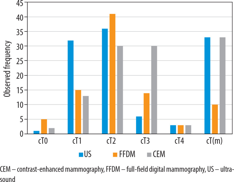
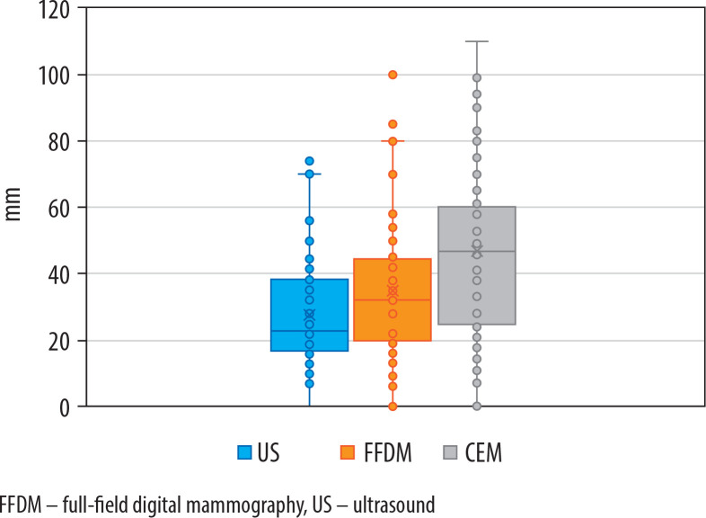
Results: Tumour size was largest in CEM (median 47 mm), followed by FFDM (median 33 mm), and smallest in US (median 23 mm). There were statistically significant differences in the distribution of cT categories between the 3 imaging modalities, with cT2 and cT1 being most common in US (46% and 41%, respectively) and FFDM (53% and 19%, respectively). Staging by CEM followed a different pattern, with cT2 and cT3 being most common (both 38%). The multiplicity rate was equal for CEM and US (42%), with fewer cases in FFDM (13%).
Conclusions: The tumour size measured by CEM is greater compared to measurements obtained through US and FFDM. Given the strong correlation between CEM and histopathology reported in the literature, CEM enhances the accuracy of local tumour staging in BC, thereby minimising the risk of understaging.

 求助内容:
求助内容: 应助结果提醒方式:


