Luca Giuliani, Nicholas Landini, Giorgio Maria Masci, Silvia Palladino, Valeria Panebianco, Giammarco Raponi, Alice Di Rocco, Giuseppe Gentile, Carlo Catalano
{"title":"血液病患者肺炎抵抗经验性治疗的ct表现。","authors":"Luca Giuliani, Nicholas Landini, Giorgio Maria Masci, Silvia Palladino, Valeria Panebianco, Giammarco Raponi, Alice Di Rocco, Giuseppe Gentile, Carlo Catalano","doi":"10.1590/0100-3984.2024.0118","DOIUrl":null,"url":null,"abstract":"<p><strong>Objective: </strong>To investigate computed tomography (CT) features of pneumonia that does not respond to empirical therapy in patients with hematologic diseases.</p><p><strong>Materials and methods: </strong>This was a retrospective analysis of all patients with hematologic disease who were diagnosed with pneumonia between 2017 and 2023, did not respond to empirical therapy for the infection, and underwent bronchoalveolar lavage and CT within a week of each other. The distribution and CT pattern of pulmonary abnormalities were assessed, as was the presence of lymphadenopathy, pleural effusion, and pericardial effusion.</p><p><strong>Results: </strong>Forty-nine patients (30 males; mean age, 61 years) were included. We identified Gram-negative bacteria in 45 patients, Gram-positive bacteria in 13, and fungi in three. Pulmonary abnormalities were bilateral in 73% of the patients in the sample, and there was no difference in prevalence between the upper and lower lung fields in 53%. Common alterations were consolidation, in 73% of the patients, bronchial wall thickening, in 71%, bronchiectasis, in 55%, and nodules, in 53%; extrapulmonary findings were less common, being identified in ≤ 27%. Pulmonary findings were typically bilateral and without a predominance between the upper and lower lung fields (<i>p</i> < 0.05). Common associations were between consolidation and bronchiectasis, between nodules and bronchial wall thickening, and between bronchiectasis and bronchial wall thickening (<i>p</i> < 0.05 for all).</p><p><strong>Conclusion: </strong>The CT manifestations of pneumonia in patients with hematologic diseases not responding to empirical therapy can resemble those of lobular pneumonia with airway inflammation. For that reason, as well as because multiple pathogens can be present in the same patient, examination of bronchoalveolar lavage fluid can be necessary.</p>","PeriodicalId":20842,"journal":{"name":"Radiologia Brasileira","volume":"58 ","pages":"e20240118"},"PeriodicalIF":0.0000,"publicationDate":"2025-07-07","publicationTypes":"Journal Article","fieldsOfStudy":null,"isOpenAccess":false,"openAccessPdf":"https://www.ncbi.nlm.nih.gov/pmc/articles/PMC12233151/pdf/","citationCount":"0","resultStr":"{\"title\":\"Computed tomography findings of pneumonia resistant to empirical therapy in patients with hematologic diseases.\",\"authors\":\"Luca Giuliani, Nicholas Landini, Giorgio Maria Masci, Silvia Palladino, Valeria Panebianco, Giammarco Raponi, Alice Di Rocco, Giuseppe Gentile, Carlo Catalano\",\"doi\":\"10.1590/0100-3984.2024.0118\",\"DOIUrl\":null,\"url\":null,\"abstract\":\"<p><strong>Objective: </strong>To investigate computed tomography (CT) features of pneumonia that does not respond to empirical therapy in patients with hematologic diseases.</p><p><strong>Materials and methods: </strong>This was a retrospective analysis of all patients with hematologic disease who were diagnosed with pneumonia between 2017 and 2023, did not respond to empirical therapy for the infection, and underwent bronchoalveolar lavage and CT within a week of each other. The distribution and CT pattern of pulmonary abnormalities were assessed, as was the presence of lymphadenopathy, pleural effusion, and pericardial effusion.</p><p><strong>Results: </strong>Forty-nine patients (30 males; mean age, 61 years) were included. We identified Gram-negative bacteria in 45 patients, Gram-positive bacteria in 13, and fungi in three. Pulmonary abnormalities were bilateral in 73% of the patients in the sample, and there was no difference in prevalence between the upper and lower lung fields in 53%. Common alterations were consolidation, in 73% of the patients, bronchial wall thickening, in 71%, bronchiectasis, in 55%, and nodules, in 53%; extrapulmonary findings were less common, being identified in ≤ 27%. Pulmonary findings were typically bilateral and without a predominance between the upper and lower lung fields (<i>p</i> < 0.05). Common associations were between consolidation and bronchiectasis, between nodules and bronchial wall thickening, and between bronchiectasis and bronchial wall thickening (<i>p</i> < 0.05 for all).</p><p><strong>Conclusion: </strong>The CT manifestations of pneumonia in patients with hematologic diseases not responding to empirical therapy can resemble those of lobular pneumonia with airway inflammation. For that reason, as well as because multiple pathogens can be present in the same patient, examination of bronchoalveolar lavage fluid can be necessary.</p>\",\"PeriodicalId\":20842,\"journal\":{\"name\":\"Radiologia Brasileira\",\"volume\":\"58 \",\"pages\":\"e20240118\"},\"PeriodicalIF\":0.0000,\"publicationDate\":\"2025-07-07\",\"publicationTypes\":\"Journal Article\",\"fieldsOfStudy\":null,\"isOpenAccess\":false,\"openAccessPdf\":\"https://www.ncbi.nlm.nih.gov/pmc/articles/PMC12233151/pdf/\",\"citationCount\":\"0\",\"resultStr\":null,\"platform\":\"Semanticscholar\",\"paperid\":null,\"PeriodicalName\":\"Radiologia Brasileira\",\"FirstCategoryId\":\"1085\",\"ListUrlMain\":\"https://doi.org/10.1590/0100-3984.2024.0118\",\"RegionNum\":0,\"RegionCategory\":null,\"ArticlePicture\":[],\"TitleCN\":null,\"AbstractTextCN\":null,\"PMCID\":null,\"EPubDate\":\"2025/1/1 0:00:00\",\"PubModel\":\"eCollection\",\"JCR\":\"Q3\",\"JCRName\":\"Medicine\",\"Score\":null,\"Total\":0}","platform":"Semanticscholar","paperid":null,"PeriodicalName":"Radiologia Brasileira","FirstCategoryId":"1085","ListUrlMain":"https://doi.org/10.1590/0100-3984.2024.0118","RegionNum":0,"RegionCategory":null,"ArticlePicture":[],"TitleCN":null,"AbstractTextCN":null,"PMCID":null,"EPubDate":"2025/1/1 0:00:00","PubModel":"eCollection","JCR":"Q3","JCRName":"Medicine","Score":null,"Total":0}
Computed tomography findings of pneumonia resistant to empirical therapy in patients with hematologic diseases.
Objective: To investigate computed tomography (CT) features of pneumonia that does not respond to empirical therapy in patients with hematologic diseases.
Materials and methods: This was a retrospective analysis of all patients with hematologic disease who were diagnosed with pneumonia between 2017 and 2023, did not respond to empirical therapy for the infection, and underwent bronchoalveolar lavage and CT within a week of each other. The distribution and CT pattern of pulmonary abnormalities were assessed, as was the presence of lymphadenopathy, pleural effusion, and pericardial effusion.
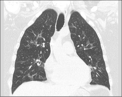
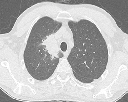
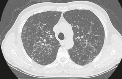
Results: Forty-nine patients (30 males; mean age, 61 years) were included. We identified Gram-negative bacteria in 45 patients, Gram-positive bacteria in 13, and fungi in three. Pulmonary abnormalities were bilateral in 73% of the patients in the sample, and there was no difference in prevalence between the upper and lower lung fields in 53%. Common alterations were consolidation, in 73% of the patients, bronchial wall thickening, in 71%, bronchiectasis, in 55%, and nodules, in 53%; extrapulmonary findings were less common, being identified in ≤ 27%. Pulmonary findings were typically bilateral and without a predominance between the upper and lower lung fields (p < 0.05). Common associations were between consolidation and bronchiectasis, between nodules and bronchial wall thickening, and between bronchiectasis and bronchial wall thickening (p < 0.05 for all).
Conclusion: The CT manifestations of pneumonia in patients with hematologic diseases not responding to empirical therapy can resemble those of lobular pneumonia with airway inflammation. For that reason, as well as because multiple pathogens can be present in the same patient, examination of bronchoalveolar lavage fluid can be necessary.

 求助内容:
求助内容: 应助结果提醒方式:


