Xiangxiang Ren, Tianhao Xie, Lingyun Liu, Fan Yang, Meng Zhang
{"title":"恶性单纯性汗腺瘤1例。","authors":"Xiangxiang Ren, Tianhao Xie, Lingyun Liu, Fan Yang, Meng Zhang","doi":"10.1159/000546700","DOIUrl":null,"url":null,"abstract":"<p><strong>Introduction: </strong>Malignant hidroacanthoma simplex (MHS) is an exceedingly rare cutaneous neoplasm with limited documented cases. This report highlights a distinctive case of MHS with prolonged clinical evolution, emphasizing its diagnostic challenges and management outcomes.</p><p><strong>Case presentation: </strong>A 64-year-old female presented with a right lumbar mass persisting for over 2 decades, exhibiting progressive enlargement in the past 5 years. Clinical examination revealed a solitary reddish-brown proliferative plaque (3.5 cm × 4.0 cm) on the right waist, characterized by irregular borders, a rough surface, and reddish-brown crusts. Histopathological findings included hyperkeratosis, irregular epidermal hyperplasia, hypertrophic stratum spinosum, and tumor cells displaying pale eosinophilic cytoplasm, vacuolated nuclei, small nucleoli, and atypical mitotic figures. Notably, tumor cells were confined to the epidermis without dermal invasion. The patient underwent local extended excision, and postoperative surveillance over 15 months demonstrated no evidence of recurrence or lymph node metastasis.</p><p><strong>Conclusion: </strong>This case underscores the indolent yet locally persistent nature of MHS. Complete surgical excision remains the cornerstone of management, with favorable outcomes achievable in the absence of dermal infiltration. Long-term follow-up is critical to monitor potential recurrence.</p>","PeriodicalId":9619,"journal":{"name":"Case Reports in Dermatology","volume":"17 1","pages":"246-251"},"PeriodicalIF":0.8000,"publicationDate":"2025-06-11","publicationTypes":"Journal Article","fieldsOfStudy":null,"isOpenAccess":false,"openAccessPdf":"https://www.ncbi.nlm.nih.gov/pmc/articles/PMC12233995/pdf/","citationCount":"0","resultStr":"{\"title\":\"Malignant Hidroacanthoma Simplex: A Case Report.\",\"authors\":\"Xiangxiang Ren, Tianhao Xie, Lingyun Liu, Fan Yang, Meng Zhang\",\"doi\":\"10.1159/000546700\",\"DOIUrl\":null,\"url\":null,\"abstract\":\"<p><strong>Introduction: </strong>Malignant hidroacanthoma simplex (MHS) is an exceedingly rare cutaneous neoplasm with limited documented cases. This report highlights a distinctive case of MHS with prolonged clinical evolution, emphasizing its diagnostic challenges and management outcomes.</p><p><strong>Case presentation: </strong>A 64-year-old female presented with a right lumbar mass persisting for over 2 decades, exhibiting progressive enlargement in the past 5 years. Clinical examination revealed a solitary reddish-brown proliferative plaque (3.5 cm × 4.0 cm) on the right waist, characterized by irregular borders, a rough surface, and reddish-brown crusts. Histopathological findings included hyperkeratosis, irregular epidermal hyperplasia, hypertrophic stratum spinosum, and tumor cells displaying pale eosinophilic cytoplasm, vacuolated nuclei, small nucleoli, and atypical mitotic figures. Notably, tumor cells were confined to the epidermis without dermal invasion. The patient underwent local extended excision, and postoperative surveillance over 15 months demonstrated no evidence of recurrence or lymph node metastasis.</p><p><strong>Conclusion: </strong>This case underscores the indolent yet locally persistent nature of MHS. Complete surgical excision remains the cornerstone of management, with favorable outcomes achievable in the absence of dermal infiltration. Long-term follow-up is critical to monitor potential recurrence.</p>\",\"PeriodicalId\":9619,\"journal\":{\"name\":\"Case Reports in Dermatology\",\"volume\":\"17 1\",\"pages\":\"246-251\"},\"PeriodicalIF\":0.8000,\"publicationDate\":\"2025-06-11\",\"publicationTypes\":\"Journal Article\",\"fieldsOfStudy\":null,\"isOpenAccess\":false,\"openAccessPdf\":\"https://www.ncbi.nlm.nih.gov/pmc/articles/PMC12233995/pdf/\",\"citationCount\":\"0\",\"resultStr\":null,\"platform\":\"Semanticscholar\",\"paperid\":null,\"PeriodicalName\":\"Case Reports in Dermatology\",\"FirstCategoryId\":\"1085\",\"ListUrlMain\":\"https://doi.org/10.1159/000546700\",\"RegionNum\":0,\"RegionCategory\":null,\"ArticlePicture\":[],\"TitleCN\":null,\"AbstractTextCN\":null,\"PMCID\":null,\"EPubDate\":\"2025/1/1 0:00:00\",\"PubModel\":\"eCollection\",\"JCR\":\"Q4\",\"JCRName\":\"DERMATOLOGY\",\"Score\":null,\"Total\":0}","platform":"Semanticscholar","paperid":null,"PeriodicalName":"Case Reports in Dermatology","FirstCategoryId":"1085","ListUrlMain":"https://doi.org/10.1159/000546700","RegionNum":0,"RegionCategory":null,"ArticlePicture":[],"TitleCN":null,"AbstractTextCN":null,"PMCID":null,"EPubDate":"2025/1/1 0:00:00","PubModel":"eCollection","JCR":"Q4","JCRName":"DERMATOLOGY","Score":null,"Total":0}
引用次数: 0
摘要
简介:恶性单纯性汗腺瘤(MHS)是一种极为罕见的皮肤肿瘤,文献记载病例有限。本报告强调了一个独特的MHS病例与长期的临床演变,强调其诊断挑战和管理结果。病例介绍:一名64岁女性,右腰椎肿块持续超过20年,在过去5年中表现出进行性扩大。临床检查示右腰一单发红褐色增生性斑块(3.5 cm × 4.0 cm),边界不规则,表面粗糙,结痂呈红褐色。组织病理学结果包括角化过度,不规则表皮增生,棘层肥大,肿瘤细胞表现为苍白嗜酸性细胞质,细胞核空泡化,核仁小,非典型有丝分裂象。值得注意的是,肿瘤细胞局限于表皮,未向真皮浸润。患者接受了局部扩大切除,术后监测超过15个月未发现复发或淋巴结转移的证据。结论:本病例强调了MHS的惰性和局部持续性。完全手术切除仍然是治疗的基石,在没有真皮浸润的情况下可以获得良好的结果。长期随访是监测潜在复发的关键。
Introduction: Malignant hidroacanthoma simplex (MHS) is an exceedingly rare cutaneous neoplasm with limited documented cases. This report highlights a distinctive case of MHS with prolonged clinical evolution, emphasizing its diagnostic challenges and management outcomes.
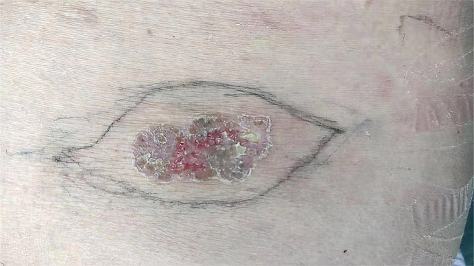
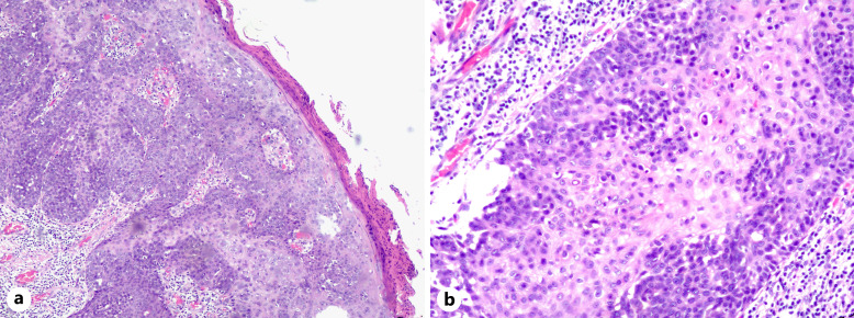
Case presentation: A 64-year-old female presented with a right lumbar mass persisting for over 2 decades, exhibiting progressive enlargement in the past 5 years. Clinical examination revealed a solitary reddish-brown proliferative plaque (3.5 cm × 4.0 cm) on the right waist, characterized by irregular borders, a rough surface, and reddish-brown crusts. Histopathological findings included hyperkeratosis, irregular epidermal hyperplasia, hypertrophic stratum spinosum, and tumor cells displaying pale eosinophilic cytoplasm, vacuolated nuclei, small nucleoli, and atypical mitotic figures. Notably, tumor cells were confined to the epidermis without dermal invasion. The patient underwent local extended excision, and postoperative surveillance over 15 months demonstrated no evidence of recurrence or lymph node metastasis.
Conclusion: This case underscores the indolent yet locally persistent nature of MHS. Complete surgical excision remains the cornerstone of management, with favorable outcomes achievable in the absence of dermal infiltration. Long-term follow-up is critical to monitor potential recurrence.

 求助内容:
求助内容: 应助结果提醒方式:


