{"title":"基于Gd-BOPTA MRI多层感知器深度学习放射组学模型识别肝细胞癌中包裹肿瘤簇的血管:一项多中心研究。","authors":"Mengting Gu, Wenjie Zou, Huilin Chen, Ruilin He, Xingyu Zhao, Ningyang Jia, Wanmin Liu, Peijun Wang","doi":"10.1186/s40644-025-00895-9","DOIUrl":null,"url":null,"abstract":"<p><strong>Objectives: </strong>The purpose of this study is to mainly develop a predictive model based on clinicoradiological and radiomics features from preoperative gadobenate-enhanced (Gd-BOPTA) magnetic resonance imaging (MRI) using multilayer perceptron (MLP) deep learning to predict vessels encapsulating tumor clusters (VETC) in hepatocellular carcinoma (HCC) patients.</p><p><strong>Methods: </strong>A total of 230 patients with histopathologically confirmed HCC who underwent preoperative Gd-BOPTA MRI before hepatectomy were retrospectively enrolled from three hospitals (144, 54, and 32 in training, test, and validation set, respectively). Univariate and multivariate logistic regression analyses were used to determine independent clinicoradiological predictors significantly associated with VETC, which then constituted the clinicoradiological model. Regions of interest (ROIs) included four modes, intratumoral (Tumor), peritumoral area ≤ 2 mm (Peri2mm), intratumoral + peritumoral area ≤ 2 mm (Tumor + Peri2mm) and intratumoral integrated with peritumoral ≤ 2 mm as a whole (TumorPeri2mm). A total of 7322 radiomics features were extracted respectively for ROI(Tumor), ROI(Peri2mm), ROI(TumorPeri2mm) and 14644 radiomics features for ROI(Tumor + Peri2mm). Least absolute shrinkage and selection operator (LASSO) and univariate logistic regression analysis were used to select the important features. Seven different machine learning classifiers respectively combined the radiomics signatures selected from four ROIs to constitute different models, and compare the performance between them in three sets and then select the optimal combination to become the radiomics model we need. Then a radiomics score (rad-score) was generated, which combined significant clinicoradiological predictors to constituted the fusion model through multivariate logistic regression analysis. After comparing the performance of the three models using area under receiver operating characteristic curve (AUC), integrated discrimination index (IDI) and net reclassification index (NRI), choose the optimal predictive model for VETC prediction.</p><p><strong>Result: </strong>Arterial peritumoral enhancement and peritumoral hypointensity on hepatobiliary phase (HBP) were independent risk factors for VETC, and constituted the Radiology model, without any clinical variables. Arterial peritumoral enhancement defined as the enhancement outside the tumor boundary in the late stage of arterial phase or early stage of portal phase, extensive contact with the tumor edge, which becomes isointense during the DP. MLP deep learning algorithm integrated radiomics features selected from ROI TumorPeri2mm was the best combination, which constituted the radiomics model (MLP model). A MLP score (MLP_score) was calculated then, which combining the two radiology features composed the fusion model (Radiology MLP model), with AUCs of 0.871, 0.894, 0.918 in the training, test and validation sets. Compared with the two models aforementioned, the Radiology MLP model demonstrated a 33.4%-131.3% improvement in NRI and a 9.3%-50% improvement in IDI, showing better discrimination, calibration and clinical usefulness in three sets, which was selected as the optimal predictive model.</p><p><strong>Conclusion: </strong>We mainly developed a fusion model (Radiology MLP model) that integrated radiology and radiomics features using MLP deep learning algorithm to predict vessels encapsulating tumor clusters (VETC) in hepatocellular carcinoma (HCC) patients, which yield an incremental value over the radiology and the MLP model.</p>","PeriodicalId":9548,"journal":{"name":"Cancer Imaging","volume":"25 1","pages":"87"},"PeriodicalIF":3.5000,"publicationDate":"2025-07-07","publicationTypes":"Journal Article","fieldsOfStudy":null,"isOpenAccess":false,"openAccessPdf":"https://www.ncbi.nlm.nih.gov/pmc/articles/PMC12232782/pdf/","citationCount":"0","resultStr":"{\"title\":\"Multilayer perceptron deep learning radiomics model based on Gd-BOPTA MRI to identify vessels encapsulating tumor clusters in hepatocellular carcinoma: a multi-center study.\",\"authors\":\"Mengting Gu, Wenjie Zou, Huilin Chen, Ruilin He, Xingyu Zhao, Ningyang Jia, Wanmin Liu, Peijun Wang\",\"doi\":\"10.1186/s40644-025-00895-9\",\"DOIUrl\":null,\"url\":null,\"abstract\":\"<p><strong>Objectives: </strong>The purpose of this study is to mainly develop a predictive model based on clinicoradiological and radiomics features from preoperative gadobenate-enhanced (Gd-BOPTA) magnetic resonance imaging (MRI) using multilayer perceptron (MLP) deep learning to predict vessels encapsulating tumor clusters (VETC) in hepatocellular carcinoma (HCC) patients.</p><p><strong>Methods: </strong>A total of 230 patients with histopathologically confirmed HCC who underwent preoperative Gd-BOPTA MRI before hepatectomy were retrospectively enrolled from three hospitals (144, 54, and 32 in training, test, and validation set, respectively). Univariate and multivariate logistic regression analyses were used to determine independent clinicoradiological predictors significantly associated with VETC, which then constituted the clinicoradiological model. Regions of interest (ROIs) included four modes, intratumoral (Tumor), peritumoral area ≤ 2 mm (Peri2mm), intratumoral + peritumoral area ≤ 2 mm (Tumor + Peri2mm) and intratumoral integrated with peritumoral ≤ 2 mm as a whole (TumorPeri2mm). A total of 7322 radiomics features were extracted respectively for ROI(Tumor), ROI(Peri2mm), ROI(TumorPeri2mm) and 14644 radiomics features for ROI(Tumor + Peri2mm). Least absolute shrinkage and selection operator (LASSO) and univariate logistic regression analysis were used to select the important features. Seven different machine learning classifiers respectively combined the radiomics signatures selected from four ROIs to constitute different models, and compare the performance between them in three sets and then select the optimal combination to become the radiomics model we need. Then a radiomics score (rad-score) was generated, which combined significant clinicoradiological predictors to constituted the fusion model through multivariate logistic regression analysis. After comparing the performance of the three models using area under receiver operating characteristic curve (AUC), integrated discrimination index (IDI) and net reclassification index (NRI), choose the optimal predictive model for VETC prediction.</p><p><strong>Result: </strong>Arterial peritumoral enhancement and peritumoral hypointensity on hepatobiliary phase (HBP) were independent risk factors for VETC, and constituted the Radiology model, without any clinical variables. Arterial peritumoral enhancement defined as the enhancement outside the tumor boundary in the late stage of arterial phase or early stage of portal phase, extensive contact with the tumor edge, which becomes isointense during the DP. MLP deep learning algorithm integrated radiomics features selected from ROI TumorPeri2mm was the best combination, which constituted the radiomics model (MLP model). A MLP score (MLP_score) was calculated then, which combining the two radiology features composed the fusion model (Radiology MLP model), with AUCs of 0.871, 0.894, 0.918 in the training, test and validation sets. Compared with the two models aforementioned, the Radiology MLP model demonstrated a 33.4%-131.3% improvement in NRI and a 9.3%-50% improvement in IDI, showing better discrimination, calibration and clinical usefulness in three sets, which was selected as the optimal predictive model.</p><p><strong>Conclusion: </strong>We mainly developed a fusion model (Radiology MLP model) that integrated radiology and radiomics features using MLP deep learning algorithm to predict vessels encapsulating tumor clusters (VETC) in hepatocellular carcinoma (HCC) patients, which yield an incremental value over the radiology and the MLP model.</p>\",\"PeriodicalId\":9548,\"journal\":{\"name\":\"Cancer Imaging\",\"volume\":\"25 1\",\"pages\":\"87\"},\"PeriodicalIF\":3.5000,\"publicationDate\":\"2025-07-07\",\"publicationTypes\":\"Journal Article\",\"fieldsOfStudy\":null,\"isOpenAccess\":false,\"openAccessPdf\":\"https://www.ncbi.nlm.nih.gov/pmc/articles/PMC12232782/pdf/\",\"citationCount\":\"0\",\"resultStr\":null,\"platform\":\"Semanticscholar\",\"paperid\":null,\"PeriodicalName\":\"Cancer Imaging\",\"FirstCategoryId\":\"3\",\"ListUrlMain\":\"https://doi.org/10.1186/s40644-025-00895-9\",\"RegionNum\":2,\"RegionCategory\":\"医学\",\"ArticlePicture\":[],\"TitleCN\":null,\"AbstractTextCN\":null,\"PMCID\":null,\"EPubDate\":\"\",\"PubModel\":\"\",\"JCR\":\"Q2\",\"JCRName\":\"ONCOLOGY\",\"Score\":null,\"Total\":0}","platform":"Semanticscholar","paperid":null,"PeriodicalName":"Cancer Imaging","FirstCategoryId":"3","ListUrlMain":"https://doi.org/10.1186/s40644-025-00895-9","RegionNum":2,"RegionCategory":"医学","ArticlePicture":[],"TitleCN":null,"AbstractTextCN":null,"PMCID":null,"EPubDate":"","PubModel":"","JCR":"Q2","JCRName":"ONCOLOGY","Score":null,"Total":0}
Multilayer perceptron deep learning radiomics model based on Gd-BOPTA MRI to identify vessels encapsulating tumor clusters in hepatocellular carcinoma: a multi-center study.
Objectives: The purpose of this study is to mainly develop a predictive model based on clinicoradiological and radiomics features from preoperative gadobenate-enhanced (Gd-BOPTA) magnetic resonance imaging (MRI) using multilayer perceptron (MLP) deep learning to predict vessels encapsulating tumor clusters (VETC) in hepatocellular carcinoma (HCC) patients.
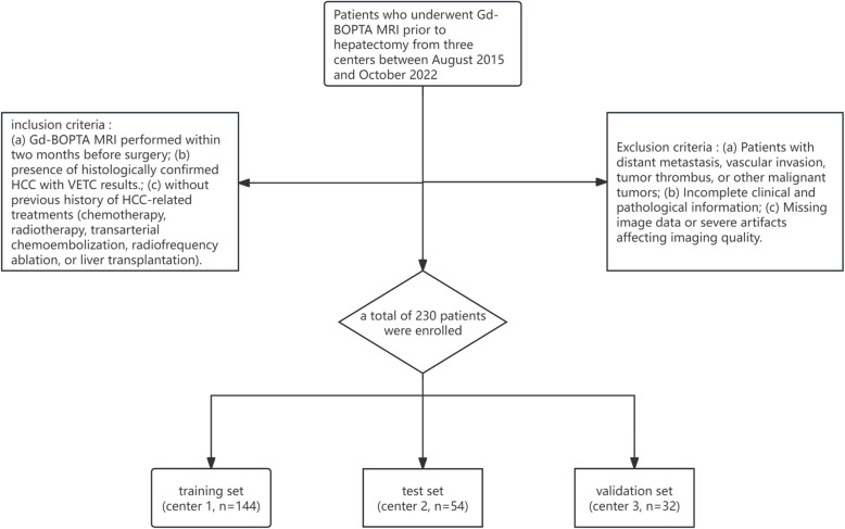
Methods: A total of 230 patients with histopathologically confirmed HCC who underwent preoperative Gd-BOPTA MRI before hepatectomy were retrospectively enrolled from three hospitals (144, 54, and 32 in training, test, and validation set, respectively). Univariate and multivariate logistic regression analyses were used to determine independent clinicoradiological predictors significantly associated with VETC, which then constituted the clinicoradiological model. Regions of interest (ROIs) included four modes, intratumoral (Tumor), peritumoral area ≤ 2 mm (Peri2mm), intratumoral + peritumoral area ≤ 2 mm (Tumor + Peri2mm) and intratumoral integrated with peritumoral ≤ 2 mm as a whole (TumorPeri2mm). A total of 7322 radiomics features were extracted respectively for ROI(Tumor), ROI(Peri2mm), ROI(TumorPeri2mm) and 14644 radiomics features for ROI(Tumor + Peri2mm). Least absolute shrinkage and selection operator (LASSO) and univariate logistic regression analysis were used to select the important features. Seven different machine learning classifiers respectively combined the radiomics signatures selected from four ROIs to constitute different models, and compare the performance between them in three sets and then select the optimal combination to become the radiomics model we need. Then a radiomics score (rad-score) was generated, which combined significant clinicoradiological predictors to constituted the fusion model through multivariate logistic regression analysis. After comparing the performance of the three models using area under receiver operating characteristic curve (AUC), integrated discrimination index (IDI) and net reclassification index (NRI), choose the optimal predictive model for VETC prediction.
Result: Arterial peritumoral enhancement and peritumoral hypointensity on hepatobiliary phase (HBP) were independent risk factors for VETC, and constituted the Radiology model, without any clinical variables. Arterial peritumoral enhancement defined as the enhancement outside the tumor boundary in the late stage of arterial phase or early stage of portal phase, extensive contact with the tumor edge, which becomes isointense during the DP. MLP deep learning algorithm integrated radiomics features selected from ROI TumorPeri2mm was the best combination, which constituted the radiomics model (MLP model). A MLP score (MLP_score) was calculated then, which combining the two radiology features composed the fusion model (Radiology MLP model), with AUCs of 0.871, 0.894, 0.918 in the training, test and validation sets. Compared with the two models aforementioned, the Radiology MLP model demonstrated a 33.4%-131.3% improvement in NRI and a 9.3%-50% improvement in IDI, showing better discrimination, calibration and clinical usefulness in three sets, which was selected as the optimal predictive model.
Conclusion: We mainly developed a fusion model (Radiology MLP model) that integrated radiology and radiomics features using MLP deep learning algorithm to predict vessels encapsulating tumor clusters (VETC) in hepatocellular carcinoma (HCC) patients, which yield an incremental value over the radiology and the MLP model.
Cancer ImagingONCOLOGY-RADIOLOGY, NUCLEAR MEDICINE & MEDICAL IMAGING
CiteScore
7.00
自引率
0.00%
发文量
66
审稿时长
>12 weeks
期刊介绍:
Cancer Imaging is an open access, peer-reviewed journal publishing original articles, reviews and editorials written by expert international radiologists working in oncology.
The journal encompasses CT, MR, PET, ultrasound, radionuclide and multimodal imaging in all kinds of malignant tumours, plus new developments, techniques and innovations. Topics of interest include:
Breast Imaging
Chest
Complications of treatment
Ear, Nose & Throat
Gastrointestinal
Hepatobiliary & Pancreatic
Imaging biomarkers
Interventional
Lymphoma
Measurement of tumour response
Molecular functional imaging
Musculoskeletal
Neuro oncology
Nuclear Medicine
Paediatric.

 求助内容:
求助内容: 应助结果提醒方式:


