{"title":"传统、半数字化和全数字化技术制备的种植支撑三单元金属框架在瓷应用前后的边缘和内部适应性","authors":"Mansour Karimi, Hamid Neshandar Asli, Yeganeh Hamrah, Mohammad Ebrahim Ghaffari, Mehran Falahchai","doi":"10.1002/cre2.70173","DOIUrl":null,"url":null,"abstract":"<div>\n \n \n <section>\n \n <h3> Objectives</h3>\n \n <p>Only a small number of studies conducted on implant-supported fixed multi-unit restorations have evaluated the semi-digital fabrication techniques. This study aimed to assess the marginal and internal adaptation of implant-supported three-unit metal frameworks fabricated by the conventional, semi-digital, and fully digital techniques before and after porcelain application.</p>\n </section>\n \n <section>\n \n <h3> Material and Methods</h3>\n \n <p>In this in vitro study, 120 three-unit metal frameworks were fabricated by five different techniques (<i>n</i> = 20): fabrication of metal frameworks from hard metal by the milling technique, direct 3D-printing of metal, milling of resin pattern and subsequent casting, 3D-printing of resin pattern and subsequent casting, and conventional waxing and subsequent casting. The marginal and internal adaptation of the frameworks was evaluated before and after porcelain application by using the silicone replica technique. Data were analyzed using ANOVA followed by pairwise comparisons with the Games-Howell and paired samples tests (α = 0.05).</p>\n </section>\n \n <section>\n \n <h3> Results</h3>\n \n <p>Before porcelain application, resin pattern milling, and subsequent casting resulted in the smallest marginal gap, while hard metal milling yielded the largest marginal gap. The fully digital techniques yielded the largest cuspal and fossa gaps, while the conventional method yielded the largest axial gap. After porcelain application, metal 3D-printing and conventional casting resulted in comparable (<i>p</i> = 0.109) marginal gaps, smaller than hard metal milling (<i>p</i> < 0.001). The conventional casting method yielded the smallest cuspal and fossa gaps (<i>p</i> < 0.001). Porcelain application significantly increased the gap size at all measurement points (<i>p</i> < 0.001).</p>\n </section>\n \n <section>\n \n <h3> Conclusion</h3>\n \n <p>The fabrication technique significantly affected the marginal and internal adaptation of implant-supported three-unit metal frameworks both before and after porcelain application.</p>\n </section>\n </div>","PeriodicalId":10203,"journal":{"name":"Clinical and Experimental Dental Research","volume":"11 4","pages":""},"PeriodicalIF":2.2000,"publicationDate":"2025-07-09","publicationTypes":"Journal Article","fieldsOfStudy":null,"isOpenAccess":false,"openAccessPdf":"https://onlinelibrary.wiley.com/doi/epdf/10.1002/cre2.70173","citationCount":"0","resultStr":"{\"title\":\"Marginal and Internal Adaptation of Implant-Supported Three-Unit Metal Frameworks Fabricated by the Conventional, Semi-Digital, and Fully Digital Techniques Before and After Porcelain Application\",\"authors\":\"Mansour Karimi, Hamid Neshandar Asli, Yeganeh Hamrah, Mohammad Ebrahim Ghaffari, Mehran Falahchai\",\"doi\":\"10.1002/cre2.70173\",\"DOIUrl\":null,\"url\":null,\"abstract\":\"<div>\\n \\n \\n <section>\\n \\n <h3> Objectives</h3>\\n \\n <p>Only a small number of studies conducted on implant-supported fixed multi-unit restorations have evaluated the semi-digital fabrication techniques. This study aimed to assess the marginal and internal adaptation of implant-supported three-unit metal frameworks fabricated by the conventional, semi-digital, and fully digital techniques before and after porcelain application.</p>\\n </section>\\n \\n <section>\\n \\n <h3> Material and Methods</h3>\\n \\n <p>In this in vitro study, 120 three-unit metal frameworks were fabricated by five different techniques (<i>n</i> = 20): fabrication of metal frameworks from hard metal by the milling technique, direct 3D-printing of metal, milling of resin pattern and subsequent casting, 3D-printing of resin pattern and subsequent casting, and conventional waxing and subsequent casting. The marginal and internal adaptation of the frameworks was evaluated before and after porcelain application by using the silicone replica technique. Data were analyzed using ANOVA followed by pairwise comparisons with the Games-Howell and paired samples tests (α = 0.05).</p>\\n </section>\\n \\n <section>\\n \\n <h3> Results</h3>\\n \\n <p>Before porcelain application, resin pattern milling, and subsequent casting resulted in the smallest marginal gap, while hard metal milling yielded the largest marginal gap. The fully digital techniques yielded the largest cuspal and fossa gaps, while the conventional method yielded the largest axial gap. After porcelain application, metal 3D-printing and conventional casting resulted in comparable (<i>p</i> = 0.109) marginal gaps, smaller than hard metal milling (<i>p</i> < 0.001). The conventional casting method yielded the smallest cuspal and fossa gaps (<i>p</i> < 0.001). Porcelain application significantly increased the gap size at all measurement points (<i>p</i> < 0.001).</p>\\n </section>\\n \\n <section>\\n \\n <h3> Conclusion</h3>\\n \\n <p>The fabrication technique significantly affected the marginal and internal adaptation of implant-supported three-unit metal frameworks both before and after porcelain application.</p>\\n </section>\\n </div>\",\"PeriodicalId\":10203,\"journal\":{\"name\":\"Clinical and Experimental Dental Research\",\"volume\":\"11 4\",\"pages\":\"\"},\"PeriodicalIF\":2.2000,\"publicationDate\":\"2025-07-09\",\"publicationTypes\":\"Journal Article\",\"fieldsOfStudy\":null,\"isOpenAccess\":false,\"openAccessPdf\":\"https://onlinelibrary.wiley.com/doi/epdf/10.1002/cre2.70173\",\"citationCount\":\"0\",\"resultStr\":null,\"platform\":\"Semanticscholar\",\"paperid\":null,\"PeriodicalName\":\"Clinical and Experimental Dental Research\",\"FirstCategoryId\":\"1085\",\"ListUrlMain\":\"https://onlinelibrary.wiley.com/doi/10.1002/cre2.70173\",\"RegionNum\":0,\"RegionCategory\":null,\"ArticlePicture\":[],\"TitleCN\":null,\"AbstractTextCN\":null,\"PMCID\":null,\"EPubDate\":\"\",\"PubModel\":\"\",\"JCR\":\"Q3\",\"JCRName\":\"DENTISTRY, ORAL SURGERY & MEDICINE\",\"Score\":null,\"Total\":0}","platform":"Semanticscholar","paperid":null,"PeriodicalName":"Clinical and Experimental Dental Research","FirstCategoryId":"1085","ListUrlMain":"https://onlinelibrary.wiley.com/doi/10.1002/cre2.70173","RegionNum":0,"RegionCategory":null,"ArticlePicture":[],"TitleCN":null,"AbstractTextCN":null,"PMCID":null,"EPubDate":"","PubModel":"","JCR":"Q3","JCRName":"DENTISTRY, ORAL SURGERY & MEDICINE","Score":null,"Total":0}
Marginal and Internal Adaptation of Implant-Supported Three-Unit Metal Frameworks Fabricated by the Conventional, Semi-Digital, and Fully Digital Techniques Before and After Porcelain Application
Objectives
Only a small number of studies conducted on implant-supported fixed multi-unit restorations have evaluated the semi-digital fabrication techniques. This study aimed to assess the marginal and internal adaptation of implant-supported three-unit metal frameworks fabricated by the conventional, semi-digital, and fully digital techniques before and after porcelain application.
Material and Methods
In this in vitro study, 120 three-unit metal frameworks were fabricated by five different techniques (n = 20): fabrication of metal frameworks from hard metal by the milling technique, direct 3D-printing of metal, milling of resin pattern and subsequent casting, 3D-printing of resin pattern and subsequent casting, and conventional waxing and subsequent casting. The marginal and internal adaptation of the frameworks was evaluated before and after porcelain application by using the silicone replica technique. Data were analyzed using ANOVA followed by pairwise comparisons with the Games-Howell and paired samples tests (α = 0.05).
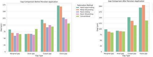
Results
Before porcelain application, resin pattern milling, and subsequent casting resulted in the smallest marginal gap, while hard metal milling yielded the largest marginal gap. The fully digital techniques yielded the largest cuspal and fossa gaps, while the conventional method yielded the largest axial gap. After porcelain application, metal 3D-printing and conventional casting resulted in comparable (p = 0.109) marginal gaps, smaller than hard metal milling (p < 0.001). The conventional casting method yielded the smallest cuspal and fossa gaps (p < 0.001). Porcelain application significantly increased the gap size at all measurement points (p < 0.001).
Conclusion
The fabrication technique significantly affected the marginal and internal adaptation of implant-supported three-unit metal frameworks both before and after porcelain application.
期刊介绍:
Clinical and Experimental Dental Research aims to provide open access peer-reviewed publications of high scientific quality representing original clinical, diagnostic or experimental work within all disciplines and fields of oral medicine and dentistry. The scope of Clinical and Experimental Dental Research comprises original research material on the anatomy, physiology and pathology of oro-facial, oro-pharyngeal and maxillofacial tissues, and functions and dysfunctions within the stomatognathic system, and the epidemiology, aetiology, prevention, diagnosis, prognosis and therapy of diseases and conditions that have an effect on the homeostasis of the mouth, jaws, and closely associated structures, as well as the healing and regeneration and the clinical aspects of replacement of hard and soft tissues with biomaterials, and the rehabilitation of stomatognathic functions. Studies that bring new knowledge on how to advance health on the individual or public health levels, including interactions between oral and general health and ill-health are welcome.

 求助内容:
求助内容: 应助结果提醒方式:


