{"title":"肌肉骨骼超声测量颈椎间盘高度的效度与信度。","authors":"Jeffrey Thompson, Jean-Michel Brismée, Phillip Page, Troy Hooper, Kathleen Rosendahl-Garcia, Stéphane Sobczak","doi":"10.26603/001c.140889","DOIUrl":null,"url":null,"abstract":"<p><strong>Background: </strong>Cervical intervertebral disc (IVD) height can be used to indirectly measure of IVD hydration status. Intervertebral disc dehydration results in height loss, which can contribute to degenerative disc disease. There is need for in situ cervical IVD ultrasound assessment to better understand spinal health.</p><p><strong>Purpose: </strong>To determine reliability and validity of musculoskeletal ultrasound (MSU) as a tool to measure cervical IVD height compared to magnetic resonance imaging (MRI) at C4-5, C5-6 and C6-7 spinal segments.</p><p><strong>Study design: </strong>Exploratory Cross-Sectional Study.</p><p><strong>Methods: </strong>This three-phase study enrolled 40 participants. Over the course of the study, 900 measurements of IVD were taken. Ten subjects participated in cervical spine MRI and MSU imaging to determine inter-rater reliability for cervical IVD height measurements. Twenty subjects underwent MRI and MSU to obtain images for measurement comparison and Bland-Altman Analysis assessed agreement between MSU and MRI (α=.05) for validity. Randomized, blinded, repeated-measures design using mean values was used to determine inter-rater reliability with intraclass correlation coefficient (ICC(2,3)) and standard error of measurement (SEM) at each IVD segment.</p><p><strong>Results: </strong>Anterior cervical IVD height of MRI and MSU were ≥0.91(95%CI=0.66-0.98) and ≥0.68(95%CI=0.27-0.92), respectively. Musculoskeletal ultrasound measurement's SEM between raters was comparable to MRI at ≤0.43mm (7.9%). No significant differences nor proportional bias between MRI and MSU measurements (p<0.05) were found at any IVD spinal level, r(18)=0.83, p<0.01. Average underestimation of MSU measurements compared to MRI was ≤ -0.10mm (2.2%).</p><p><strong>Conclusion: </strong>Methodology used for MSU cervical IVD height imaging and measurements was found to be moderately to highly reliable. Comparisons measurements between MRI and MSU support the use of MSU to measure cervical IVD height in future investigations, including variables which may affect the IVD hydration and homeostasis.</p><p><strong>Level of evidence: </strong>I 3.</p>","PeriodicalId":47892,"journal":{"name":"International Journal of Sports Physical Therapy","volume":"20 7","pages":"964-973"},"PeriodicalIF":2.1000,"publicationDate":"2025-07-02","publicationTypes":"Journal Article","fieldsOfStudy":null,"isOpenAccess":false,"openAccessPdf":"https://www.ncbi.nlm.nih.gov/pmc/articles/PMC12222554/pdf/","citationCount":"0","resultStr":"{\"title\":\"Validity & Reliability of Using Musculoskeletal Ultrasound to Measure Cervical Disc Height.\",\"authors\":\"Jeffrey Thompson, Jean-Michel Brismée, Phillip Page, Troy Hooper, Kathleen Rosendahl-Garcia, Stéphane Sobczak\",\"doi\":\"10.26603/001c.140889\",\"DOIUrl\":null,\"url\":null,\"abstract\":\"<p><strong>Background: </strong>Cervical intervertebral disc (IVD) height can be used to indirectly measure of IVD hydration status. Intervertebral disc dehydration results in height loss, which can contribute to degenerative disc disease. There is need for in situ cervical IVD ultrasound assessment to better understand spinal health.</p><p><strong>Purpose: </strong>To determine reliability and validity of musculoskeletal ultrasound (MSU) as a tool to measure cervical IVD height compared to magnetic resonance imaging (MRI) at C4-5, C5-6 and C6-7 spinal segments.</p><p><strong>Study design: </strong>Exploratory Cross-Sectional Study.</p><p><strong>Methods: </strong>This three-phase study enrolled 40 participants. Over the course of the study, 900 measurements of IVD were taken. Ten subjects participated in cervical spine MRI and MSU imaging to determine inter-rater reliability for cervical IVD height measurements. Twenty subjects underwent MRI and MSU to obtain images for measurement comparison and Bland-Altman Analysis assessed agreement between MSU and MRI (α=.05) for validity. Randomized, blinded, repeated-measures design using mean values was used to determine inter-rater reliability with intraclass correlation coefficient (ICC(2,3)) and standard error of measurement (SEM) at each IVD segment.</p><p><strong>Results: </strong>Anterior cervical IVD height of MRI and MSU were ≥0.91(95%CI=0.66-0.98) and ≥0.68(95%CI=0.27-0.92), respectively. Musculoskeletal ultrasound measurement's SEM between raters was comparable to MRI at ≤0.43mm (7.9%). No significant differences nor proportional bias between MRI and MSU measurements (p<0.05) were found at any IVD spinal level, r(18)=0.83, p<0.01. Average underestimation of MSU measurements compared to MRI was ≤ -0.10mm (2.2%).</p><p><strong>Conclusion: </strong>Methodology used for MSU cervical IVD height imaging and measurements was found to be moderately to highly reliable. Comparisons measurements between MRI and MSU support the use of MSU to measure cervical IVD height in future investigations, including variables which may affect the IVD hydration and homeostasis.</p><p><strong>Level of evidence: </strong>I 3.</p>\",\"PeriodicalId\":47892,\"journal\":{\"name\":\"International Journal of Sports Physical Therapy\",\"volume\":\"20 7\",\"pages\":\"964-973\"},\"PeriodicalIF\":2.1000,\"publicationDate\":\"2025-07-02\",\"publicationTypes\":\"Journal Article\",\"fieldsOfStudy\":null,\"isOpenAccess\":false,\"openAccessPdf\":\"https://www.ncbi.nlm.nih.gov/pmc/articles/PMC12222554/pdf/\",\"citationCount\":\"0\",\"resultStr\":null,\"platform\":\"Semanticscholar\",\"paperid\":null,\"PeriodicalName\":\"International Journal of Sports Physical Therapy\",\"FirstCategoryId\":\"1085\",\"ListUrlMain\":\"https://doi.org/10.26603/001c.140889\",\"RegionNum\":0,\"RegionCategory\":null,\"ArticlePicture\":[],\"TitleCN\":null,\"AbstractTextCN\":null,\"PMCID\":null,\"EPubDate\":\"2025/1/1 0:00:00\",\"PubModel\":\"eCollection\",\"JCR\":\"Q3\",\"JCRName\":\"SPORT SCIENCES\",\"Score\":null,\"Total\":0}","platform":"Semanticscholar","paperid":null,"PeriodicalName":"International Journal of Sports Physical Therapy","FirstCategoryId":"1085","ListUrlMain":"https://doi.org/10.26603/001c.140889","RegionNum":0,"RegionCategory":null,"ArticlePicture":[],"TitleCN":null,"AbstractTextCN":null,"PMCID":null,"EPubDate":"2025/1/1 0:00:00","PubModel":"eCollection","JCR":"Q3","JCRName":"SPORT SCIENCES","Score":null,"Total":0}
Validity & Reliability of Using Musculoskeletal Ultrasound to Measure Cervical Disc Height.
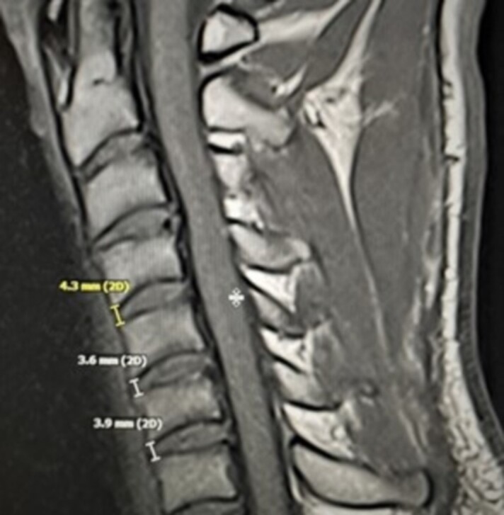
Background: Cervical intervertebral disc (IVD) height can be used to indirectly measure of IVD hydration status. Intervertebral disc dehydration results in height loss, which can contribute to degenerative disc disease. There is need for in situ cervical IVD ultrasound assessment to better understand spinal health.
Purpose: To determine reliability and validity of musculoskeletal ultrasound (MSU) as a tool to measure cervical IVD height compared to magnetic resonance imaging (MRI) at C4-5, C5-6 and C6-7 spinal segments.
Study design: Exploratory Cross-Sectional Study.
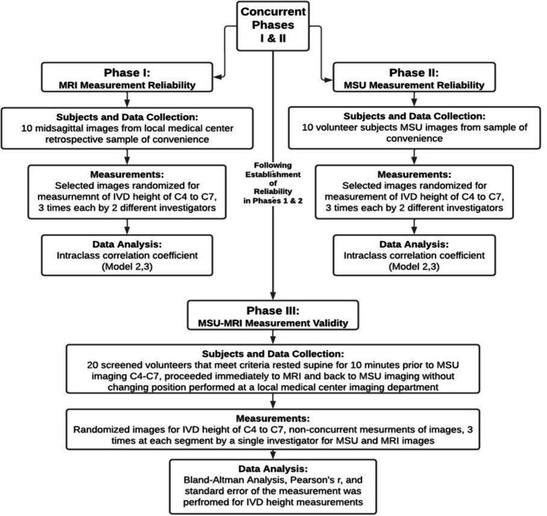
Methods: This three-phase study enrolled 40 participants. Over the course of the study, 900 measurements of IVD were taken. Ten subjects participated in cervical spine MRI and MSU imaging to determine inter-rater reliability for cervical IVD height measurements. Twenty subjects underwent MRI and MSU to obtain images for measurement comparison and Bland-Altman Analysis assessed agreement between MSU and MRI (α=.05) for validity. Randomized, blinded, repeated-measures design using mean values was used to determine inter-rater reliability with intraclass correlation coefficient (ICC(2,3)) and standard error of measurement (SEM) at each IVD segment.
Results: Anterior cervical IVD height of MRI and MSU were ≥0.91(95%CI=0.66-0.98) and ≥0.68(95%CI=0.27-0.92), respectively. Musculoskeletal ultrasound measurement's SEM between raters was comparable to MRI at ≤0.43mm (7.9%). No significant differences nor proportional bias between MRI and MSU measurements (p<0.05) were found at any IVD spinal level, r(18)=0.83, p<0.01. Average underestimation of MSU measurements compared to MRI was ≤ -0.10mm (2.2%).
Conclusion: Methodology used for MSU cervical IVD height imaging and measurements was found to be moderately to highly reliable. Comparisons measurements between MRI and MSU support the use of MSU to measure cervical IVD height in future investigations, including variables which may affect the IVD hydration and homeostasis.


 求助内容:
求助内容: 应助结果提醒方式:


