{"title":"牙釉质厚度三维评价指导正畸近端间复位:一项基于cbct的跨性别和种族研究。","authors":"Ezgi Cansu Fırıncıoğulları, Aslıhan Ertan Erdinç, Sercan Akyalçın","doi":"10.4274/TurkJOrthod.2025.2025.36","DOIUrl":null,"url":null,"abstract":"<p><strong>Objective: </strong>This study aimed to explore variations in enamel thickness to provide guidelines for optimal interproximal enamel reduction in an untreated population using cone-beam computed tomography (CBCT).</p><p><strong>Methods: </strong>CBCT scans of 100 orthodontic patients (51 Caucasian, 49 patients of Somalian descent; aged (12-18) were analyzed retrospectively. Enamel thickness was measured at the mesial and distal contact points of teeth from the second molar to the central incisor in both the maxillary and mandibular arches. Linear mixed models were employed to assess the effects of ethnicity, gender, anterior-posterior region, and mesial-distal proximal surfaces on enamel thickness. Fixed effects were estimated using the Kenward-Roger method, and a random intercept with an unstructured covariance matrix was included to account for within-subject variability. Ethnicity-specific residual variances were also modeled. Statistical significance was set at p<0.05.</p><p><strong>Results: </strong>Enamel thickness varied significantly between Caucasians and Somalians in both the maxilla and mandible (p<0.001), with greater thickness observed in Caucasians. Gender-related differences were minimal; however, in the maxilla, distal surfaces of posterior teeth had greater enamel thickness in females compared to males (p=0.0478). Enamel thickness was consistently greater on distal surfaces of posterior teeth (p<0.001), while no significant differences were observed between mesial and distal surfaces in anterior teeth (p>0.05).</p><p><strong>Conclusion: </strong>Posterior teeth, particularly distal proximal surfaces of premolars and molars hold a great potential for enamel reduction, offering clinicians the most optimal site in orthodontic interventions.</p>","PeriodicalId":37013,"journal":{"name":"Turkish Journal of Orthodontics","volume":"38 2","pages":"89-96"},"PeriodicalIF":1.4000,"publicationDate":"2025-07-02","publicationTypes":"Journal Article","fieldsOfStudy":null,"isOpenAccess":false,"openAccessPdf":"https://www.ncbi.nlm.nih.gov/pmc/articles/PMC12236122/pdf/","citationCount":"0","resultStr":"{\"title\":\"3-Dimensional Evaluation of Enamel Thickness to Guide Orthodontic Interproximal Reduction: A CBCT-Based Study Across Gender and Ethnicity.\",\"authors\":\"Ezgi Cansu Fırıncıoğulları, Aslıhan Ertan Erdinç, Sercan Akyalçın\",\"doi\":\"10.4274/TurkJOrthod.2025.2025.36\",\"DOIUrl\":null,\"url\":null,\"abstract\":\"<p><strong>Objective: </strong>This study aimed to explore variations in enamel thickness to provide guidelines for optimal interproximal enamel reduction in an untreated population using cone-beam computed tomography (CBCT).</p><p><strong>Methods: </strong>CBCT scans of 100 orthodontic patients (51 Caucasian, 49 patients of Somalian descent; aged (12-18) were analyzed retrospectively. Enamel thickness was measured at the mesial and distal contact points of teeth from the second molar to the central incisor in both the maxillary and mandibular arches. Linear mixed models were employed to assess the effects of ethnicity, gender, anterior-posterior region, and mesial-distal proximal surfaces on enamel thickness. Fixed effects were estimated using the Kenward-Roger method, and a random intercept with an unstructured covariance matrix was included to account for within-subject variability. Ethnicity-specific residual variances were also modeled. Statistical significance was set at p<0.05.</p><p><strong>Results: </strong>Enamel thickness varied significantly between Caucasians and Somalians in both the maxilla and mandible (p<0.001), with greater thickness observed in Caucasians. Gender-related differences were minimal; however, in the maxilla, distal surfaces of posterior teeth had greater enamel thickness in females compared to males (p=0.0478). Enamel thickness was consistently greater on distal surfaces of posterior teeth (p<0.001), while no significant differences were observed between mesial and distal surfaces in anterior teeth (p>0.05).</p><p><strong>Conclusion: </strong>Posterior teeth, particularly distal proximal surfaces of premolars and molars hold a great potential for enamel reduction, offering clinicians the most optimal site in orthodontic interventions.</p>\",\"PeriodicalId\":37013,\"journal\":{\"name\":\"Turkish Journal of Orthodontics\",\"volume\":\"38 2\",\"pages\":\"89-96\"},\"PeriodicalIF\":1.4000,\"publicationDate\":\"2025-07-02\",\"publicationTypes\":\"Journal Article\",\"fieldsOfStudy\":null,\"isOpenAccess\":false,\"openAccessPdf\":\"https://www.ncbi.nlm.nih.gov/pmc/articles/PMC12236122/pdf/\",\"citationCount\":\"0\",\"resultStr\":null,\"platform\":\"Semanticscholar\",\"paperid\":null,\"PeriodicalName\":\"Turkish Journal of Orthodontics\",\"FirstCategoryId\":\"1085\",\"ListUrlMain\":\"https://doi.org/10.4274/TurkJOrthod.2025.2025.36\",\"RegionNum\":0,\"RegionCategory\":null,\"ArticlePicture\":[],\"TitleCN\":null,\"AbstractTextCN\":null,\"PMCID\":null,\"EPubDate\":\"\",\"PubModel\":\"\",\"JCR\":\"Q4\",\"JCRName\":\"DENTISTRY, ORAL SURGERY & MEDICINE\",\"Score\":null,\"Total\":0}","platform":"Semanticscholar","paperid":null,"PeriodicalName":"Turkish Journal of Orthodontics","FirstCategoryId":"1085","ListUrlMain":"https://doi.org/10.4274/TurkJOrthod.2025.2025.36","RegionNum":0,"RegionCategory":null,"ArticlePicture":[],"TitleCN":null,"AbstractTextCN":null,"PMCID":null,"EPubDate":"","PubModel":"","JCR":"Q4","JCRName":"DENTISTRY, ORAL SURGERY & MEDICINE","Score":null,"Total":0}
3-Dimensional Evaluation of Enamel Thickness to Guide Orthodontic Interproximal Reduction: A CBCT-Based Study Across Gender and Ethnicity.
Objective: This study aimed to explore variations in enamel thickness to provide guidelines for optimal interproximal enamel reduction in an untreated population using cone-beam computed tomography (CBCT).
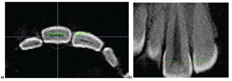
Methods: CBCT scans of 100 orthodontic patients (51 Caucasian, 49 patients of Somalian descent; aged (12-18) were analyzed retrospectively. Enamel thickness was measured at the mesial and distal contact points of teeth from the second molar to the central incisor in both the maxillary and mandibular arches. Linear mixed models were employed to assess the effects of ethnicity, gender, anterior-posterior region, and mesial-distal proximal surfaces on enamel thickness. Fixed effects were estimated using the Kenward-Roger method, and a random intercept with an unstructured covariance matrix was included to account for within-subject variability. Ethnicity-specific residual variances were also modeled. Statistical significance was set at p<0.05.
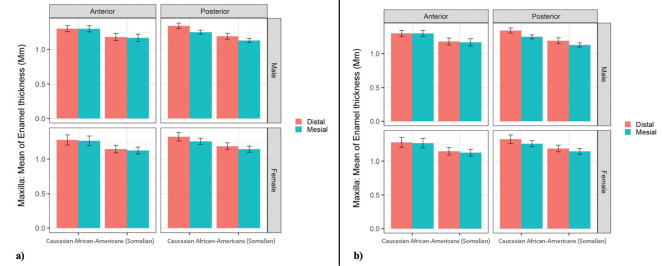
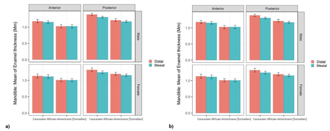
Results: Enamel thickness varied significantly between Caucasians and Somalians in both the maxilla and mandible (p<0.001), with greater thickness observed in Caucasians. Gender-related differences were minimal; however, in the maxilla, distal surfaces of posterior teeth had greater enamel thickness in females compared to males (p=0.0478). Enamel thickness was consistently greater on distal surfaces of posterior teeth (p<0.001), while no significant differences were observed between mesial and distal surfaces in anterior teeth (p>0.05).
Conclusion: Posterior teeth, particularly distal proximal surfaces of premolars and molars hold a great potential for enamel reduction, offering clinicians the most optimal site in orthodontic interventions.

 求助内容:
求助内容: 应助结果提醒方式:


