Parkavi Arumugam, Bianca Princeton, Pradeep Kumar Yadalam, Carlos M Ardila
{"title":"创新羟基磷灰石涂层二氧化钛纳米管用于牙种植体表面增强。","authors":"Parkavi Arumugam, Bianca Princeton, Pradeep Kumar Yadalam, Carlos M Ardila","doi":"10.4317/jced.62626","DOIUrl":null,"url":null,"abstract":"<p><strong>Background: </strong>The present study aimed to develop novel hydroxyapatite-coated titanium dioxide or titania nanotubes (TNTs) as a surface modification on titanium dental implants and analyze their surface, chemical properties, biocompatibility, and corrosion resistance.</p><p><strong>Material and methods: </strong>The titanium implant surface was treated with 1 ml of Kroll's reagent, 5 ml of nitric acid, 1.5 ml of sulfuric acid, and water for 10 seconds to allow etching of the surface. The etched surface was then anodized to create a layer of titanium dioxide, which, on treatment with 1wt% of hydrofluoric acid in water under the anodization process with 100 volts for 1 hour at room temperature, led to the formation of TNTs. The nanotube surface was then dipped in Hank's solution, allowing hydroxyapatite deposition on the surface. After 7 days, the hydroxyapatite-coated TNTs (GROUP A) as a surface coating on titanium implants was characterized and compared with bare titanium implants (Group B).</p><p><strong>Results: </strong>The material characterization showed successful development of hydroxyapatite-coated TNT formation on titanium implant surface, which supported cell adhesion, proliferation, and migration, similar to uncoated titanium surfaces. No statistically significant difference in the percentage of cell viability was noted between Groups A and B at any time point, with the highest percentage of cell viability with a mean of 93.20 +/- 4.324 for Group A and 94.00 +/- 6.205 for Group B noted at 72 hours, with a p-value of 0.21. Corrosion testing showed the coating's higher corrosion potential and reduced corrosion density compared to uncoated titanium surfaces with the bode phase angle approaching 1, suggesting its potential for better clinical outcomes.</p><p><strong>Conclusions: </strong>The hydroxyapatite-coated TNTs have good surface, chemical corrosion-resistant properties, and optimal biocompatibility. Further in vivo studies are warranted to assess the osteogenic and antimicrobial properties, as well as the clinical efficacy, of this coating. <b>Key words:</b>Dental implant, hydroxyapatite, titania nanotubes, biocompatibility, corrosion, osseointegration.</p>","PeriodicalId":15376,"journal":{"name":"Journal of Clinical and Experimental Dentistry","volume":"17 6","pages":"e695-e704"},"PeriodicalIF":0.0000,"publicationDate":"2025-06-01","publicationTypes":"Journal Article","fieldsOfStudy":null,"isOpenAccess":false,"openAccessPdf":"https://www.ncbi.nlm.nih.gov/pmc/articles/PMC12225762/pdf/","citationCount":"0","resultStr":"{\"title\":\"Innovative Hydroxyapatite-Coated Titania Nanotubes for Dental Implant Surface Enhancement.\",\"authors\":\"Parkavi Arumugam, Bianca Princeton, Pradeep Kumar Yadalam, Carlos M Ardila\",\"doi\":\"10.4317/jced.62626\",\"DOIUrl\":null,\"url\":null,\"abstract\":\"<p><strong>Background: </strong>The present study aimed to develop novel hydroxyapatite-coated titanium dioxide or titania nanotubes (TNTs) as a surface modification on titanium dental implants and analyze their surface, chemical properties, biocompatibility, and corrosion resistance.</p><p><strong>Material and methods: </strong>The titanium implant surface was treated with 1 ml of Kroll's reagent, 5 ml of nitric acid, 1.5 ml of sulfuric acid, and water for 10 seconds to allow etching of the surface. The etched surface was then anodized to create a layer of titanium dioxide, which, on treatment with 1wt% of hydrofluoric acid in water under the anodization process with 100 volts for 1 hour at room temperature, led to the formation of TNTs. The nanotube surface was then dipped in Hank's solution, allowing hydroxyapatite deposition on the surface. After 7 days, the hydroxyapatite-coated TNTs (GROUP A) as a surface coating on titanium implants was characterized and compared with bare titanium implants (Group B).</p><p><strong>Results: </strong>The material characterization showed successful development of hydroxyapatite-coated TNT formation on titanium implant surface, which supported cell adhesion, proliferation, and migration, similar to uncoated titanium surfaces. No statistically significant difference in the percentage of cell viability was noted between Groups A and B at any time point, with the highest percentage of cell viability with a mean of 93.20 +/- 4.324 for Group A and 94.00 +/- 6.205 for Group B noted at 72 hours, with a p-value of 0.21. Corrosion testing showed the coating's higher corrosion potential and reduced corrosion density compared to uncoated titanium surfaces with the bode phase angle approaching 1, suggesting its potential for better clinical outcomes.</p><p><strong>Conclusions: </strong>The hydroxyapatite-coated TNTs have good surface, chemical corrosion-resistant properties, and optimal biocompatibility. Further in vivo studies are warranted to assess the osteogenic and antimicrobial properties, as well as the clinical efficacy, of this coating. <b>Key words:</b>Dental implant, hydroxyapatite, titania nanotubes, biocompatibility, corrosion, osseointegration.</p>\",\"PeriodicalId\":15376,\"journal\":{\"name\":\"Journal of Clinical and Experimental Dentistry\",\"volume\":\"17 6\",\"pages\":\"e695-e704\"},\"PeriodicalIF\":0.0000,\"publicationDate\":\"2025-06-01\",\"publicationTypes\":\"Journal Article\",\"fieldsOfStudy\":null,\"isOpenAccess\":false,\"openAccessPdf\":\"https://www.ncbi.nlm.nih.gov/pmc/articles/PMC12225762/pdf/\",\"citationCount\":\"0\",\"resultStr\":null,\"platform\":\"Semanticscholar\",\"paperid\":null,\"PeriodicalName\":\"Journal of Clinical and Experimental Dentistry\",\"FirstCategoryId\":\"1085\",\"ListUrlMain\":\"https://doi.org/10.4317/jced.62626\",\"RegionNum\":0,\"RegionCategory\":null,\"ArticlePicture\":[],\"TitleCN\":null,\"AbstractTextCN\":null,\"PMCID\":null,\"EPubDate\":\"\",\"PubModel\":\"\",\"JCR\":\"Q2\",\"JCRName\":\"Dentistry\",\"Score\":null,\"Total\":0}","platform":"Semanticscholar","paperid":null,"PeriodicalName":"Journal of Clinical and Experimental Dentistry","FirstCategoryId":"1085","ListUrlMain":"https://doi.org/10.4317/jced.62626","RegionNum":0,"RegionCategory":null,"ArticlePicture":[],"TitleCN":null,"AbstractTextCN":null,"PMCID":null,"EPubDate":"","PubModel":"","JCR":"Q2","JCRName":"Dentistry","Score":null,"Total":0}
Innovative Hydroxyapatite-Coated Titania Nanotubes for Dental Implant Surface Enhancement.
Background: The present study aimed to develop novel hydroxyapatite-coated titanium dioxide or titania nanotubes (TNTs) as a surface modification on titanium dental implants and analyze their surface, chemical properties, biocompatibility, and corrosion resistance.
Material and methods: The titanium implant surface was treated with 1 ml of Kroll's reagent, 5 ml of nitric acid, 1.5 ml of sulfuric acid, and water for 10 seconds to allow etching of the surface. The etched surface was then anodized to create a layer of titanium dioxide, which, on treatment with 1wt% of hydrofluoric acid in water under the anodization process with 100 volts for 1 hour at room temperature, led to the formation of TNTs. The nanotube surface was then dipped in Hank's solution, allowing hydroxyapatite deposition on the surface. After 7 days, the hydroxyapatite-coated TNTs (GROUP A) as a surface coating on titanium implants was characterized and compared with bare titanium implants (Group B).
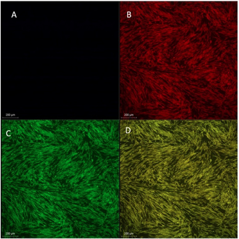
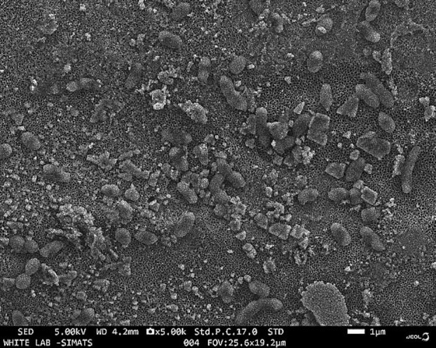
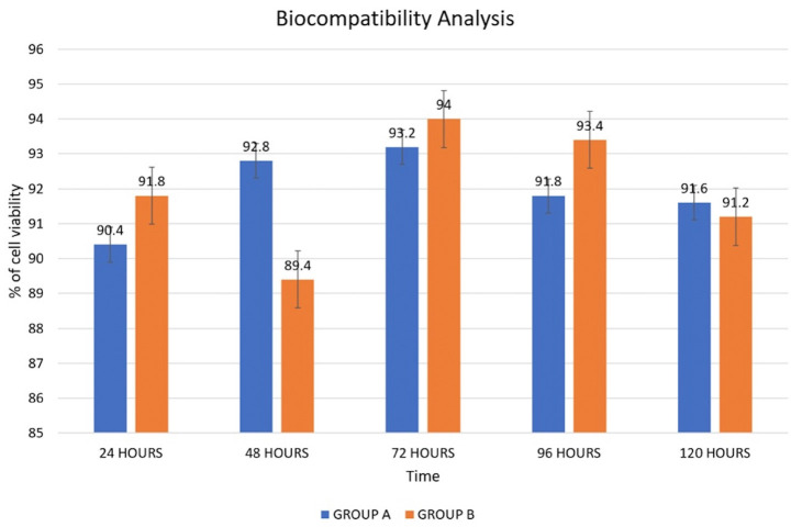
Results: The material characterization showed successful development of hydroxyapatite-coated TNT formation on titanium implant surface, which supported cell adhesion, proliferation, and migration, similar to uncoated titanium surfaces. No statistically significant difference in the percentage of cell viability was noted between Groups A and B at any time point, with the highest percentage of cell viability with a mean of 93.20 +/- 4.324 for Group A and 94.00 +/- 6.205 for Group B noted at 72 hours, with a p-value of 0.21. Corrosion testing showed the coating's higher corrosion potential and reduced corrosion density compared to uncoated titanium surfaces with the bode phase angle approaching 1, suggesting its potential for better clinical outcomes.
Conclusions: The hydroxyapatite-coated TNTs have good surface, chemical corrosion-resistant properties, and optimal biocompatibility. Further in vivo studies are warranted to assess the osteogenic and antimicrobial properties, as well as the clinical efficacy, of this coating. Key words:Dental implant, hydroxyapatite, titania nanotubes, biocompatibility, corrosion, osseointegration.
期刊介绍:
Indexed in PUBMED, PubMed Central® (PMC) since 2012 and SCOPUSJournal of Clinical and Experimental Dentistry is an Open Access (free access on-line) - http://www.medicinaoral.com/odo/indice.htm. The aim of the Journal of Clinical and Experimental Dentistry is: - Periodontology - Community and Preventive Dentistry - Esthetic Dentistry - Biomaterials and Bioengineering in Dentistry - Operative Dentistry and Endodontics - Prosthetic Dentistry - Orthodontics - Oral Medicine and Pathology - Odontostomatology for the disabled or special patients - Oral Surgery

 求助内容:
求助内容: 应助结果提醒方式:


