Lu Jin, Meng Ding, Shaoxin Cui, Lin Yang, Jinwen Zhao, Jingjing He, Xiaoping Wang, Fei Chang, Xue Liu, Qun Wang, Hongtao Jin, Jun Ma, Aijing Liu
{"title":"异常糖代谢驱动系统性红斑狼疮CD4+ T细胞功能障碍和疾病爆发。","authors":"Lu Jin, Meng Ding, Shaoxin Cui, Lin Yang, Jinwen Zhao, Jingjing He, Xiaoping Wang, Fei Chang, Xue Liu, Qun Wang, Hongtao Jin, Jun Ma, Aijing Liu","doi":"10.5114/ceji.2025.149252","DOIUrl":null,"url":null,"abstract":"<p><strong>Introduction: </strong>T cell immuno-metabolic regulation plays a key role in the development of systemic lupus erythematosus (SLE). This study aimed to analyze the role of CD4<sup>+</sup> T cell glucose metabolism in SLE development.</p><p><strong>Material and methods: </strong>Clinical data and blood samples were collected from 20 untreated SLE patients and healthy controls (HCs) matched for age, sex, and body mass index. After being isolated by magnetic sorting and cultured with anti-CD3/CD28 for 72 h, CD4<sup>+</sup> T cells were subjected to real-time metabolic analysis. CD4<sup>+</sup> T cell proliferation and cytokines were measured with cell counting kit-8 and Luminex liquid chip assay, respectively.</p><p><strong>Results: </strong>Compared to HCs, SLE-CD4<sup>+</sup> T cells exhibited significantly higher glycolytic capacity and mitochondrial oxidative phosphorylation (OXPHOS) (both p < 0.001). Additionally, SLE-CD4<sup>+</sup> T cells demonstrated increased proliferation rates and elevated cytokine levels in both plasma and culture supernatants (both p < 0.05). OXPHOS and glycolysis of SLE-CD4<sup>+</sup> T cells were positively correlated with SLE disease activity index-2000 (SLEDAI-2K) and cytokines, and negatively correlated with SLE-CD4<sup>+</sup> T cell numbers (all p < 0.05).</p><p><strong>Conclusions: </strong>CD4<sup>+</sup> T cells from SLE patients showed higher glucose metabolic activity than those from HCs, and the enhanced glucose metabolism of SLE-CD4<sup>+</sup> T cells was strongly correlated with disease activity, suggesting that glucose metabolic reprogramming plays an essential role in the pathogenesis of SLE.</p>","PeriodicalId":9694,"journal":{"name":"Central European Journal of Immunology","volume":"50 1","pages":"13-23"},"PeriodicalIF":1.6000,"publicationDate":"2025-01-01","publicationTypes":"Journal Article","fieldsOfStudy":null,"isOpenAccess":false,"openAccessPdf":"https://www.ncbi.nlm.nih.gov/pmc/articles/PMC12224248/pdf/","citationCount":"0","resultStr":"{\"title\":\"Aberrant glucose metabolism drives dysfunction of CD4<sup>+</sup> T cells in systemic lupus erythematosus and disease flares.\",\"authors\":\"Lu Jin, Meng Ding, Shaoxin Cui, Lin Yang, Jinwen Zhao, Jingjing He, Xiaoping Wang, Fei Chang, Xue Liu, Qun Wang, Hongtao Jin, Jun Ma, Aijing Liu\",\"doi\":\"10.5114/ceji.2025.149252\",\"DOIUrl\":null,\"url\":null,\"abstract\":\"<p><strong>Introduction: </strong>T cell immuno-metabolic regulation plays a key role in the development of systemic lupus erythematosus (SLE). This study aimed to analyze the role of CD4<sup>+</sup> T cell glucose metabolism in SLE development.</p><p><strong>Material and methods: </strong>Clinical data and blood samples were collected from 20 untreated SLE patients and healthy controls (HCs) matched for age, sex, and body mass index. After being isolated by magnetic sorting and cultured with anti-CD3/CD28 for 72 h, CD4<sup>+</sup> T cells were subjected to real-time metabolic analysis. CD4<sup>+</sup> T cell proliferation and cytokines were measured with cell counting kit-8 and Luminex liquid chip assay, respectively.</p><p><strong>Results: </strong>Compared to HCs, SLE-CD4<sup>+</sup> T cells exhibited significantly higher glycolytic capacity and mitochondrial oxidative phosphorylation (OXPHOS) (both p < 0.001). Additionally, SLE-CD4<sup>+</sup> T cells demonstrated increased proliferation rates and elevated cytokine levels in both plasma and culture supernatants (both p < 0.05). OXPHOS and glycolysis of SLE-CD4<sup>+</sup> T cells were positively correlated with SLE disease activity index-2000 (SLEDAI-2K) and cytokines, and negatively correlated with SLE-CD4<sup>+</sup> T cell numbers (all p < 0.05).</p><p><strong>Conclusions: </strong>CD4<sup>+</sup> T cells from SLE patients showed higher glucose metabolic activity than those from HCs, and the enhanced glucose metabolism of SLE-CD4<sup>+</sup> T cells was strongly correlated with disease activity, suggesting that glucose metabolic reprogramming plays an essential role in the pathogenesis of SLE.</p>\",\"PeriodicalId\":9694,\"journal\":{\"name\":\"Central European Journal of Immunology\",\"volume\":\"50 1\",\"pages\":\"13-23\"},\"PeriodicalIF\":1.6000,\"publicationDate\":\"2025-01-01\",\"publicationTypes\":\"Journal Article\",\"fieldsOfStudy\":null,\"isOpenAccess\":false,\"openAccessPdf\":\"https://www.ncbi.nlm.nih.gov/pmc/articles/PMC12224248/pdf/\",\"citationCount\":\"0\",\"resultStr\":null,\"platform\":\"Semanticscholar\",\"paperid\":null,\"PeriodicalName\":\"Central European Journal of Immunology\",\"FirstCategoryId\":\"3\",\"ListUrlMain\":\"https://doi.org/10.5114/ceji.2025.149252\",\"RegionNum\":4,\"RegionCategory\":\"医学\",\"ArticlePicture\":[],\"TitleCN\":null,\"AbstractTextCN\":null,\"PMCID\":null,\"EPubDate\":\"2025/4/9 0:00:00\",\"PubModel\":\"Epub\",\"JCR\":\"Q4\",\"JCRName\":\"IMMUNOLOGY\",\"Score\":null,\"Total\":0}","platform":"Semanticscholar","paperid":null,"PeriodicalName":"Central European Journal of Immunology","FirstCategoryId":"3","ListUrlMain":"https://doi.org/10.5114/ceji.2025.149252","RegionNum":4,"RegionCategory":"医学","ArticlePicture":[],"TitleCN":null,"AbstractTextCN":null,"PMCID":null,"EPubDate":"2025/4/9 0:00:00","PubModel":"Epub","JCR":"Q4","JCRName":"IMMUNOLOGY","Score":null,"Total":0}
Aberrant glucose metabolism drives dysfunction of CD4+ T cells in systemic lupus erythematosus and disease flares.
Introduction: T cell immuno-metabolic regulation plays a key role in the development of systemic lupus erythematosus (SLE). This study aimed to analyze the role of CD4+ T cell glucose metabolism in SLE development.
Material and methods: Clinical data and blood samples were collected from 20 untreated SLE patients and healthy controls (HCs) matched for age, sex, and body mass index. After being isolated by magnetic sorting and cultured with anti-CD3/CD28 for 72 h, CD4+ T cells were subjected to real-time metabolic analysis. CD4+ T cell proliferation and cytokines were measured with cell counting kit-8 and Luminex liquid chip assay, respectively.
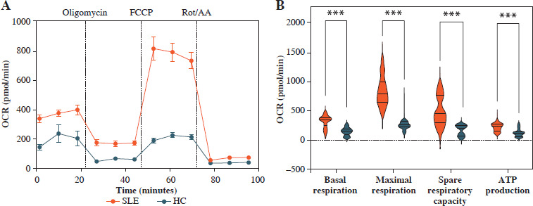
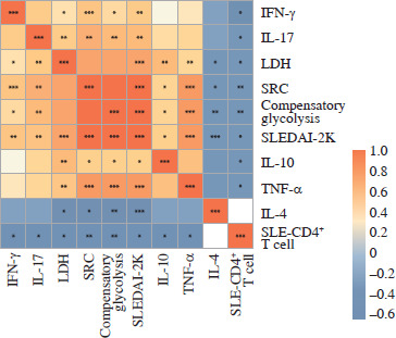
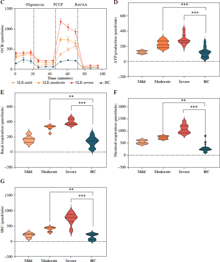
Results: Compared to HCs, SLE-CD4+ T cells exhibited significantly higher glycolytic capacity and mitochondrial oxidative phosphorylation (OXPHOS) (both p < 0.001). Additionally, SLE-CD4+ T cells demonstrated increased proliferation rates and elevated cytokine levels in both plasma and culture supernatants (both p < 0.05). OXPHOS and glycolysis of SLE-CD4+ T cells were positively correlated with SLE disease activity index-2000 (SLEDAI-2K) and cytokines, and negatively correlated with SLE-CD4+ T cell numbers (all p < 0.05).
Conclusions: CD4+ T cells from SLE patients showed higher glucose metabolic activity than those from HCs, and the enhanced glucose metabolism of SLE-CD4+ T cells was strongly correlated with disease activity, suggesting that glucose metabolic reprogramming plays an essential role in the pathogenesis of SLE.
 求助内容:
求助内容: 应助结果提醒方式:


