Esther Bankole, Chun Wai Wong, Sally Kim, Matthew Hind, Charlotte H Dean
{"title":"人PCLS肺损伤及修复模型的发现及药物研究。","authors":"Esther Bankole, Chun Wai Wong, Sally Kim, Matthew Hind, Charlotte H Dean","doi":"10.1186/s12931-025-03314-6","DOIUrl":null,"url":null,"abstract":"<p><strong>Background: </strong>The Acid Injury and Repair (AIR) model is an ex-vivo model of lung injury and repair, that was previously established using mouse precision-cut lung slices (PCLS). The AIR model provides a bridge between the current in -vitro and in-vivo models to study the effects of lung injury in 3D lung tissue slices. Here, we show that the AIR model can be adapted for use in human tissue as a translational model for discovery research and drug screening.</p><p><strong>Methods: </strong>To generate PCLS, resected human lung tissue was coated with alginate hydrogel to form an artificial pleura. Lung tissue was inflated by point injecting 3% agarose, followed by generation of 450-500 µM thick slices of tissue. An isolated area of each slice was injured by brief application of 0.1 M hydrochloric acid. AIR-PCLS were then washed and cultured for 48 h before immunostaining to assess proliferating cells (Ki67) alveolar type II/progenitor cell markers (HTII, proSP-C), lipofibroblasts (ADRP) and endothelial cells (ERG). Viability of PCLS was assessed by both MTT assay and Live/Dead staining.</p><p><strong>Results: </strong>We show that levels of proliferation do not change in response to acid injury. However, there is a significant increase in the percentage of proSP-C and HTII positive cells in the injured regions of AIR-PCLS. We also identify non-epithelial cell populations; lipofibroblasts and endothelial cells in human AIR-PCLS, to demonstrate that other repair relevant cell types can be identified and tracked in the human AIR (hAIR model).</p><p><strong>Conclusions: </strong>The hAIR model is an effective ex-vivo tool to study early mechanisms of lung repair following injury. By establishing an area of injured tissue adjacent to uninjured tissue, this model mimics the heterogenous pattern of lung injury frequently present in lung diseases. The hAIR model will facilitate mechanistic studies of human lung repair and provides a valuable pre-clinical model for drug testing.</p>","PeriodicalId":49131,"journal":{"name":"Respiratory Research","volume":"26 1","pages":"237"},"PeriodicalIF":5.8000,"publicationDate":"2025-07-05","publicationTypes":"Journal Article","fieldsOfStudy":null,"isOpenAccess":false,"openAccessPdf":"https://www.ncbi.nlm.nih.gov/pmc/articles/PMC12228282/pdf/","citationCount":"0","resultStr":"{\"title\":\"A human PCLS model of lung injury and repair for discovery and pharmaceutical research.\",\"authors\":\"Esther Bankole, Chun Wai Wong, Sally Kim, Matthew Hind, Charlotte H Dean\",\"doi\":\"10.1186/s12931-025-03314-6\",\"DOIUrl\":null,\"url\":null,\"abstract\":\"<p><strong>Background: </strong>The Acid Injury and Repair (AIR) model is an ex-vivo model of lung injury and repair, that was previously established using mouse precision-cut lung slices (PCLS). The AIR model provides a bridge between the current in -vitro and in-vivo models to study the effects of lung injury in 3D lung tissue slices. Here, we show that the AIR model can be adapted for use in human tissue as a translational model for discovery research and drug screening.</p><p><strong>Methods: </strong>To generate PCLS, resected human lung tissue was coated with alginate hydrogel to form an artificial pleura. Lung tissue was inflated by point injecting 3% agarose, followed by generation of 450-500 µM thick slices of tissue. An isolated area of each slice was injured by brief application of 0.1 M hydrochloric acid. AIR-PCLS were then washed and cultured for 48 h before immunostaining to assess proliferating cells (Ki67) alveolar type II/progenitor cell markers (HTII, proSP-C), lipofibroblasts (ADRP) and endothelial cells (ERG). Viability of PCLS was assessed by both MTT assay and Live/Dead staining.</p><p><strong>Results: </strong>We show that levels of proliferation do not change in response to acid injury. However, there is a significant increase in the percentage of proSP-C and HTII positive cells in the injured regions of AIR-PCLS. We also identify non-epithelial cell populations; lipofibroblasts and endothelial cells in human AIR-PCLS, to demonstrate that other repair relevant cell types can be identified and tracked in the human AIR (hAIR model).</p><p><strong>Conclusions: </strong>The hAIR model is an effective ex-vivo tool to study early mechanisms of lung repair following injury. By establishing an area of injured tissue adjacent to uninjured tissue, this model mimics the heterogenous pattern of lung injury frequently present in lung diseases. The hAIR model will facilitate mechanistic studies of human lung repair and provides a valuable pre-clinical model for drug testing.</p>\",\"PeriodicalId\":49131,\"journal\":{\"name\":\"Respiratory Research\",\"volume\":\"26 1\",\"pages\":\"237\"},\"PeriodicalIF\":5.8000,\"publicationDate\":\"2025-07-05\",\"publicationTypes\":\"Journal Article\",\"fieldsOfStudy\":null,\"isOpenAccess\":false,\"openAccessPdf\":\"https://www.ncbi.nlm.nih.gov/pmc/articles/PMC12228282/pdf/\",\"citationCount\":\"0\",\"resultStr\":null,\"platform\":\"Semanticscholar\",\"paperid\":null,\"PeriodicalName\":\"Respiratory Research\",\"FirstCategoryId\":\"3\",\"ListUrlMain\":\"https://doi.org/10.1186/s12931-025-03314-6\",\"RegionNum\":2,\"RegionCategory\":\"医学\",\"ArticlePicture\":[],\"TitleCN\":null,\"AbstractTextCN\":null,\"PMCID\":null,\"EPubDate\":\"\",\"PubModel\":\"\",\"JCR\":\"Q1\",\"JCRName\":\"Medicine\",\"Score\":null,\"Total\":0}","platform":"Semanticscholar","paperid":null,"PeriodicalName":"Respiratory Research","FirstCategoryId":"3","ListUrlMain":"https://doi.org/10.1186/s12931-025-03314-6","RegionNum":2,"RegionCategory":"医学","ArticlePicture":[],"TitleCN":null,"AbstractTextCN":null,"PMCID":null,"EPubDate":"","PubModel":"","JCR":"Q1","JCRName":"Medicine","Score":null,"Total":0}
引用次数: 0
摘要
背景:酸性损伤与修复(AIR)模型是一种离体肺损伤与修复模型,先前使用小鼠精确切割肺切片(PCLS)建立。AIR模型是目前体外和体内模型之间的桥梁,可以在三维肺组织切片中研究肺损伤的影响。在这里,我们展示了AIR模型可以适用于人体组织,作为发现研究和药物筛选的转化模型。方法:采用海藻酸盐水凝胶包被人肺组织形成人工胸膜,制备PCLS。点注射3%琼脂糖膨胀肺组织,生成450 ~ 500µM厚的肺组织切片。用0.1 M盐酸对每片切片的孤立区域进行短暂的损伤。然后将AIR-PCLS洗涤并培养48小时,然后进行免疫染色,以评估增殖细胞(Ki67)肺泡II型/祖细胞标志物(HTII, pro - c),脂肪成纤维细胞(ADRP)和内皮细胞(ERG)。采用MTT法和活/死染色法评估PCLS的生存能力。结果:我们表明,在酸损伤的反应中,增殖水平没有改变。然而,在AIR-PCLS损伤区域中,pro - c和HTII阳性细胞的百分比显著增加。我们还鉴定了非上皮细胞群;人类AIR- pcls中的脂肪成纤维细胞和内皮细胞,以证明在人类AIR(毛发模型)中可以识别和追踪其他与修复相关的细胞类型。结论:毛发模型是研究损伤后肺修复早期机制的有效离体工具。通过建立一个与未损伤组织相邻的损伤组织区域,该模型模拟了肺部疾病中常见的肺损伤的异质性模式。hAIR模型将促进人体肺修复的机制研究,并为药物测试提供有价值的临床前模型。
A human PCLS model of lung injury and repair for discovery and pharmaceutical research.
Background: The Acid Injury and Repair (AIR) model is an ex-vivo model of lung injury and repair, that was previously established using mouse precision-cut lung slices (PCLS). The AIR model provides a bridge between the current in -vitro and in-vivo models to study the effects of lung injury in 3D lung tissue slices. Here, we show that the AIR model can be adapted for use in human tissue as a translational model for discovery research and drug screening.
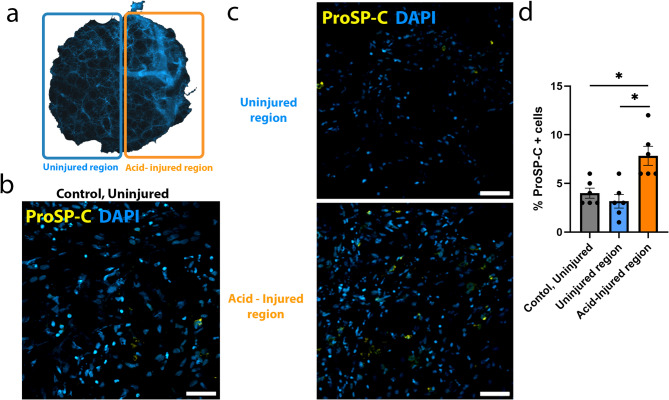
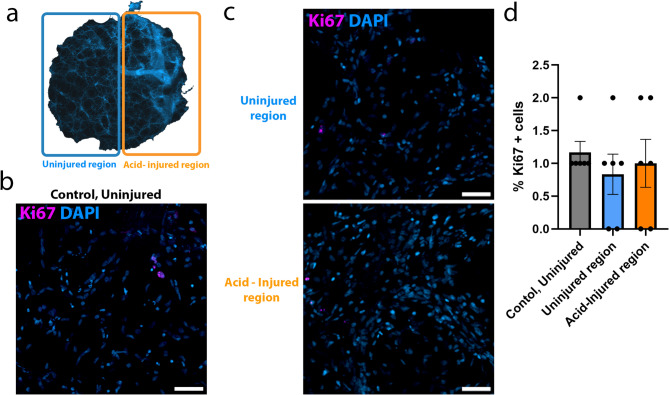
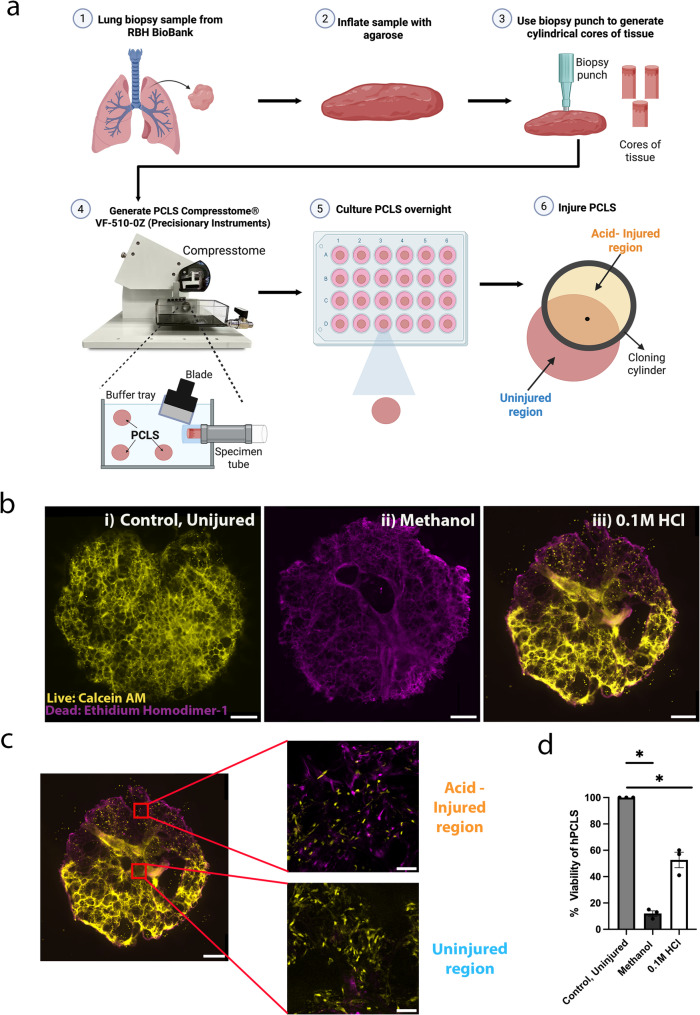
Methods: To generate PCLS, resected human lung tissue was coated with alginate hydrogel to form an artificial pleura. Lung tissue was inflated by point injecting 3% agarose, followed by generation of 450-500 µM thick slices of tissue. An isolated area of each slice was injured by brief application of 0.1 M hydrochloric acid. AIR-PCLS were then washed and cultured for 48 h before immunostaining to assess proliferating cells (Ki67) alveolar type II/progenitor cell markers (HTII, proSP-C), lipofibroblasts (ADRP) and endothelial cells (ERG). Viability of PCLS was assessed by both MTT assay and Live/Dead staining.
Results: We show that levels of proliferation do not change in response to acid injury. However, there is a significant increase in the percentage of proSP-C and HTII positive cells in the injured regions of AIR-PCLS. We also identify non-epithelial cell populations; lipofibroblasts and endothelial cells in human AIR-PCLS, to demonstrate that other repair relevant cell types can be identified and tracked in the human AIR (hAIR model).
Conclusions: The hAIR model is an effective ex-vivo tool to study early mechanisms of lung repair following injury. By establishing an area of injured tissue adjacent to uninjured tissue, this model mimics the heterogenous pattern of lung injury frequently present in lung diseases. The hAIR model will facilitate mechanistic studies of human lung repair and provides a valuable pre-clinical model for drug testing.
期刊介绍:
Respiratory Research publishes high-quality clinical and basic research, review and commentary articles on all aspects of respiratory medicine and related diseases.
As the leading fully open access journal in the field, Respiratory Research provides an essential resource for pulmonologists, allergists, immunologists and other physicians, researchers, healthcare workers and medical students with worldwide dissemination of articles resulting in high visibility and generating international discussion.
Topics of specific interest include asthma, chronic obstructive pulmonary disease, cystic fibrosis, genetics, infectious diseases, interstitial lung diseases, lung development, lung tumors, occupational and environmental factors, pulmonary circulation, pulmonary pharmacology and therapeutics, respiratory immunology, respiratory physiology, and sleep-related respiratory problems.
 求助内容:
求助内容: 应助结果提醒方式:


