Eric Toshiyuki Nakamura, Francisco Tustumi, Marina Alessandra Pereira, Leonardo Cardili, Ana Paula Gárate, Bruno Cogliati, Venancio Avancini Ferreira Alves, Sandra Nassa Sampietre, Cinthia Lanchotte Ferreira, Giovanna Mattos Ferreira, Michelly Moreira Campos, Ulysses Ribeiro Junior, Luiz Augusto Carneiro D'Albuquerque, Flávio Henrique Ferreira Galvão
{"title":"预测切口疝复发的可重复大鼠模型:对临床翻译的见解。","authors":"Eric Toshiyuki Nakamura, Francisco Tustumi, Marina Alessandra Pereira, Leonardo Cardili, Ana Paula Gárate, Bruno Cogliati, Venancio Avancini Ferreira Alves, Sandra Nassa Sampietre, Cinthia Lanchotte Ferreira, Giovanna Mattos Ferreira, Michelly Moreira Campos, Ulysses Ribeiro Junior, Luiz Augusto Carneiro D'Albuquerque, Flávio Henrique Ferreira Galvão","doi":"10.1038/s41598-025-05557-1","DOIUrl":null,"url":null,"abstract":"<p><p>Incisional hernias are common complications following abdominal surgeries. This study aimed to develop a reproducible murine model of complex incisional hernia to better understand this condition and evaluate new therapeutic strategies. Fourteen male Wistar rats underwent laparotomy to induce hernia formation. Intra-abdominal volume was measured. Hernia recurrence, tissue healing, and adhesion formation were evaluated through macroscopic and histopathological analysis, assessing fibrosis, angiogenesis, inflammation, and necrosis. All rats developed incisional hernias after laparotomy, with a 90% recurrence rate observed one week post-repair. The average hernia defect size was 10.30 mm ± 9.32 mm. A significant 25.63% reduction in intra-abdominal volume was recorded. Macroscopic examination revealed adhesions in 80% of the animals, with 60% classified as severe. Histopathological analysis showed fibrosis in all animals, with 70% displaying moderate to severe fibrosis, characterized by multifocal areas of recent fibrosis or signs of myofibroblastic differentiation. Inflammation, indicated by granulation tissue, was present in all animals. Necrosis was observed in 60% of the animals. Fibrosis affected 40% of the incision areas and 70% of the abdominal muscles. This animal model has proven versatile, reproducible, and reliable, making it suitable for investigating complex incisional hernias in translational studies.</p>","PeriodicalId":21811,"journal":{"name":"Scientific Reports","volume":"15 1","pages":"23861"},"PeriodicalIF":3.9000,"publicationDate":"2025-07-04","publicationTypes":"Journal Article","fieldsOfStudy":null,"isOpenAccess":false,"openAccessPdf":"https://www.ncbi.nlm.nih.gov/pmc/articles/PMC12227744/pdf/","citationCount":"0","resultStr":"{\"title\":\"A reproducible rat model for predicting incisional hernia recurrence: insights for clinical translation.\",\"authors\":\"Eric Toshiyuki Nakamura, Francisco Tustumi, Marina Alessandra Pereira, Leonardo Cardili, Ana Paula Gárate, Bruno Cogliati, Venancio Avancini Ferreira Alves, Sandra Nassa Sampietre, Cinthia Lanchotte Ferreira, Giovanna Mattos Ferreira, Michelly Moreira Campos, Ulysses Ribeiro Junior, Luiz Augusto Carneiro D'Albuquerque, Flávio Henrique Ferreira Galvão\",\"doi\":\"10.1038/s41598-025-05557-1\",\"DOIUrl\":null,\"url\":null,\"abstract\":\"<p><p>Incisional hernias are common complications following abdominal surgeries. This study aimed to develop a reproducible murine model of complex incisional hernia to better understand this condition and evaluate new therapeutic strategies. Fourteen male Wistar rats underwent laparotomy to induce hernia formation. Intra-abdominal volume was measured. Hernia recurrence, tissue healing, and adhesion formation were evaluated through macroscopic and histopathological analysis, assessing fibrosis, angiogenesis, inflammation, and necrosis. All rats developed incisional hernias after laparotomy, with a 90% recurrence rate observed one week post-repair. The average hernia defect size was 10.30 mm ± 9.32 mm. A significant 25.63% reduction in intra-abdominal volume was recorded. Macroscopic examination revealed adhesions in 80% of the animals, with 60% classified as severe. Histopathological analysis showed fibrosis in all animals, with 70% displaying moderate to severe fibrosis, characterized by multifocal areas of recent fibrosis or signs of myofibroblastic differentiation. Inflammation, indicated by granulation tissue, was present in all animals. Necrosis was observed in 60% of the animals. Fibrosis affected 40% of the incision areas and 70% of the abdominal muscles. This animal model has proven versatile, reproducible, and reliable, making it suitable for investigating complex incisional hernias in translational studies.</p>\",\"PeriodicalId\":21811,\"journal\":{\"name\":\"Scientific Reports\",\"volume\":\"15 1\",\"pages\":\"23861\"},\"PeriodicalIF\":3.9000,\"publicationDate\":\"2025-07-04\",\"publicationTypes\":\"Journal Article\",\"fieldsOfStudy\":null,\"isOpenAccess\":false,\"openAccessPdf\":\"https://www.ncbi.nlm.nih.gov/pmc/articles/PMC12227744/pdf/\",\"citationCount\":\"0\",\"resultStr\":null,\"platform\":\"Semanticscholar\",\"paperid\":null,\"PeriodicalName\":\"Scientific Reports\",\"FirstCategoryId\":\"103\",\"ListUrlMain\":\"https://doi.org/10.1038/s41598-025-05557-1\",\"RegionNum\":2,\"RegionCategory\":\"综合性期刊\",\"ArticlePicture\":[],\"TitleCN\":null,\"AbstractTextCN\":null,\"PMCID\":null,\"EPubDate\":\"\",\"PubModel\":\"\",\"JCR\":\"Q1\",\"JCRName\":\"MULTIDISCIPLINARY SCIENCES\",\"Score\":null,\"Total\":0}","platform":"Semanticscholar","paperid":null,"PeriodicalName":"Scientific Reports","FirstCategoryId":"103","ListUrlMain":"https://doi.org/10.1038/s41598-025-05557-1","RegionNum":2,"RegionCategory":"综合性期刊","ArticlePicture":[],"TitleCN":null,"AbstractTextCN":null,"PMCID":null,"EPubDate":"","PubModel":"","JCR":"Q1","JCRName":"MULTIDISCIPLINARY SCIENCES","Score":null,"Total":0}
A reproducible rat model for predicting incisional hernia recurrence: insights for clinical translation.
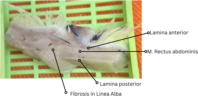
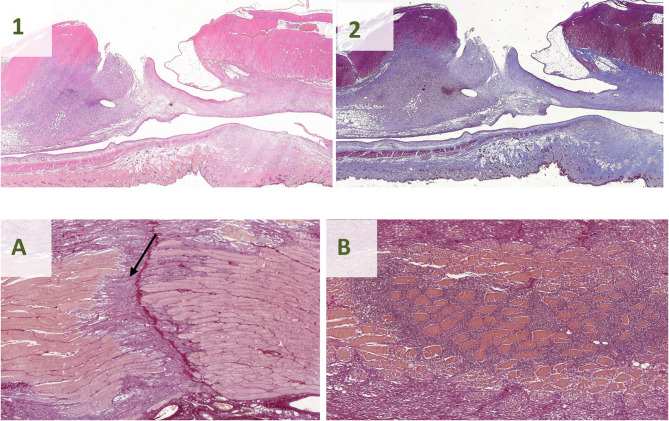
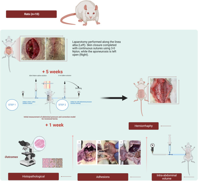
Incisional hernias are common complications following abdominal surgeries. This study aimed to develop a reproducible murine model of complex incisional hernia to better understand this condition and evaluate new therapeutic strategies. Fourteen male Wistar rats underwent laparotomy to induce hernia formation. Intra-abdominal volume was measured. Hernia recurrence, tissue healing, and adhesion formation were evaluated through macroscopic and histopathological analysis, assessing fibrosis, angiogenesis, inflammation, and necrosis. All rats developed incisional hernias after laparotomy, with a 90% recurrence rate observed one week post-repair. The average hernia defect size was 10.30 mm ± 9.32 mm. A significant 25.63% reduction in intra-abdominal volume was recorded. Macroscopic examination revealed adhesions in 80% of the animals, with 60% classified as severe. Histopathological analysis showed fibrosis in all animals, with 70% displaying moderate to severe fibrosis, characterized by multifocal areas of recent fibrosis or signs of myofibroblastic differentiation. Inflammation, indicated by granulation tissue, was present in all animals. Necrosis was observed in 60% of the animals. Fibrosis affected 40% of the incision areas and 70% of the abdominal muscles. This animal model has proven versatile, reproducible, and reliable, making it suitable for investigating complex incisional hernias in translational studies.
期刊介绍:
We publish original research from all areas of the natural sciences, psychology, medicine and engineering. You can learn more about what we publish by browsing our specific scientific subject areas below or explore Scientific Reports by browsing all articles and collections.
Scientific Reports has a 2-year impact factor: 4.380 (2021), and is the 6th most-cited journal in the world, with more than 540,000 citations in 2020 (Clarivate Analytics, 2021).
•Engineering
Engineering covers all aspects of engineering, technology, and applied science. It plays a crucial role in the development of technologies to address some of the world''s biggest challenges, helping to save lives and improve the way we live.
•Physical sciences
Physical sciences are those academic disciplines that aim to uncover the underlying laws of nature — often written in the language of mathematics. It is a collective term for areas of study including astronomy, chemistry, materials science and physics.
•Earth and environmental sciences
Earth and environmental sciences cover all aspects of Earth and planetary science and broadly encompass solid Earth processes, surface and atmospheric dynamics, Earth system history, climate and climate change, marine and freshwater systems, and ecology. It also considers the interactions between humans and these systems.
•Biological sciences
Biological sciences encompass all the divisions of natural sciences examining various aspects of vital processes. The concept includes anatomy, physiology, cell biology, biochemistry and biophysics, and covers all organisms from microorganisms, animals to plants.
•Health sciences
The health sciences study health, disease and healthcare. This field of study aims to develop knowledge, interventions and technology for use in healthcare to improve the treatment of patients.
 求助内容:
求助内容: 应助结果提醒方式:


