Ammar Almarghlani, Reem A Alsahafi, Fatimah K Alqahtani, Yousef Alnowailaty, Mohammed Barayan, Ameerah Aladwani, Amr Bokhari
{"title":"使用CBCT评估牙周炎患者的牙髓变化:体积分析。","authors":"Ammar Almarghlani, Reem A Alsahafi, Fatimah K Alqahtani, Yousef Alnowailaty, Mohammed Barayan, Ameerah Aladwani, Amr Bokhari","doi":"10.3389/fdmed.2025.1549281","DOIUrl":null,"url":null,"abstract":"<p><strong>Introduction: </strong>Evidence suggests that periodontal disease can lead to inflammation and degeneration within dental pulp, highlighting the need for dental professionals to understand this association better.</p><p><strong>Objective: </strong>The objective for this study was to establish a correlation between pulp volume and periodontal disease using Cone-Beam Computed Tomography (CBCT) imaging.</p><p><strong>Methods: </strong>A cross-sectional study design was employed for the collected data from 148 patients aged 39.51 years using dental images obtained by CBCT and analyzed by medical software to create three-dimensional (3D) images. Pulp-volume analysis was performed using Amira software and measurements were derived using bio-models generated from CBCT images. Obtained data was analyzed using SPSS-27 statistical software.</p><p><strong>Results: </strong>The mean pulp volume between healthy and teeth with periodontitis showed certain differences. The mean low and largest pulp volumes of 9.15 ± 3.3 mm<sup>3</sup> and 15.24 ± 4.2 mm<sup>3</sup> were observed involving teeth # 41 and teeth # 13, respectively. Furthermore, a higher mean of pulp volume was observed in healthy teeth than in periodontitis-diagnosed teeth except for teeth # 33 and 43. The significant difference (<i>p</i> < 0.05) was easily detected involving teeth # 22, 23, 11, and 13. However, the lowest difference, with non-significant difference (<i>p</i> > 0.05), involving teeth # 43, 31, and 33 was observed.</p><p><strong>Discussion: </strong>The study's findings underscore a significant correlation between periodontitis and reduced pulp volume, suggesting that periodontal inflammation may influence pupal remodeling.</p>","PeriodicalId":73077,"journal":{"name":"Frontiers in dental medicine","volume":"6 ","pages":"1549281"},"PeriodicalIF":1.8000,"publicationDate":"2025-06-19","publicationTypes":"Journal Article","fieldsOfStudy":null,"isOpenAccess":false,"openAccessPdf":"https://www.ncbi.nlm.nih.gov/pmc/articles/PMC12222284/pdf/","citationCount":"0","resultStr":"{\"title\":\"Assessment of pulpal changes in periodontitis patients using CBCT: a volumetric analysis.\",\"authors\":\"Ammar Almarghlani, Reem A Alsahafi, Fatimah K Alqahtani, Yousef Alnowailaty, Mohammed Barayan, Ameerah Aladwani, Amr Bokhari\",\"doi\":\"10.3389/fdmed.2025.1549281\",\"DOIUrl\":null,\"url\":null,\"abstract\":\"<p><strong>Introduction: </strong>Evidence suggests that periodontal disease can lead to inflammation and degeneration within dental pulp, highlighting the need for dental professionals to understand this association better.</p><p><strong>Objective: </strong>The objective for this study was to establish a correlation between pulp volume and periodontal disease using Cone-Beam Computed Tomography (CBCT) imaging.</p><p><strong>Methods: </strong>A cross-sectional study design was employed for the collected data from 148 patients aged 39.51 years using dental images obtained by CBCT and analyzed by medical software to create three-dimensional (3D) images. Pulp-volume analysis was performed using Amira software and measurements were derived using bio-models generated from CBCT images. Obtained data was analyzed using SPSS-27 statistical software.</p><p><strong>Results: </strong>The mean pulp volume between healthy and teeth with periodontitis showed certain differences. The mean low and largest pulp volumes of 9.15 ± 3.3 mm<sup>3</sup> and 15.24 ± 4.2 mm<sup>3</sup> were observed involving teeth # 41 and teeth # 13, respectively. Furthermore, a higher mean of pulp volume was observed in healthy teeth than in periodontitis-diagnosed teeth except for teeth # 33 and 43. The significant difference (<i>p</i> < 0.05) was easily detected involving teeth # 22, 23, 11, and 13. However, the lowest difference, with non-significant difference (<i>p</i> > 0.05), involving teeth # 43, 31, and 33 was observed.</p><p><strong>Discussion: </strong>The study's findings underscore a significant correlation between periodontitis and reduced pulp volume, suggesting that periodontal inflammation may influence pupal remodeling.</p>\",\"PeriodicalId\":73077,\"journal\":{\"name\":\"Frontiers in dental medicine\",\"volume\":\"6 \",\"pages\":\"1549281\"},\"PeriodicalIF\":1.8000,\"publicationDate\":\"2025-06-19\",\"publicationTypes\":\"Journal Article\",\"fieldsOfStudy\":null,\"isOpenAccess\":false,\"openAccessPdf\":\"https://www.ncbi.nlm.nih.gov/pmc/articles/PMC12222284/pdf/\",\"citationCount\":\"0\",\"resultStr\":null,\"platform\":\"Semanticscholar\",\"paperid\":null,\"PeriodicalName\":\"Frontiers in dental medicine\",\"FirstCategoryId\":\"1085\",\"ListUrlMain\":\"https://doi.org/10.3389/fdmed.2025.1549281\",\"RegionNum\":0,\"RegionCategory\":null,\"ArticlePicture\":[],\"TitleCN\":null,\"AbstractTextCN\":null,\"PMCID\":null,\"EPubDate\":\"2025/1/1 0:00:00\",\"PubModel\":\"eCollection\",\"JCR\":\"Q3\",\"JCRName\":\"DENTISTRY, ORAL SURGERY & MEDICINE\",\"Score\":null,\"Total\":0}","platform":"Semanticscholar","paperid":null,"PeriodicalName":"Frontiers in dental medicine","FirstCategoryId":"1085","ListUrlMain":"https://doi.org/10.3389/fdmed.2025.1549281","RegionNum":0,"RegionCategory":null,"ArticlePicture":[],"TitleCN":null,"AbstractTextCN":null,"PMCID":null,"EPubDate":"2025/1/1 0:00:00","PubModel":"eCollection","JCR":"Q3","JCRName":"DENTISTRY, ORAL SURGERY & MEDICINE","Score":null,"Total":0}
Assessment of pulpal changes in periodontitis patients using CBCT: a volumetric analysis.
Introduction: Evidence suggests that periodontal disease can lead to inflammation and degeneration within dental pulp, highlighting the need for dental professionals to understand this association better.
Objective: The objective for this study was to establish a correlation between pulp volume and periodontal disease using Cone-Beam Computed Tomography (CBCT) imaging.
Methods: A cross-sectional study design was employed for the collected data from 148 patients aged 39.51 years using dental images obtained by CBCT and analyzed by medical software to create three-dimensional (3D) images. Pulp-volume analysis was performed using Amira software and measurements were derived using bio-models generated from CBCT images. Obtained data was analyzed using SPSS-27 statistical software.
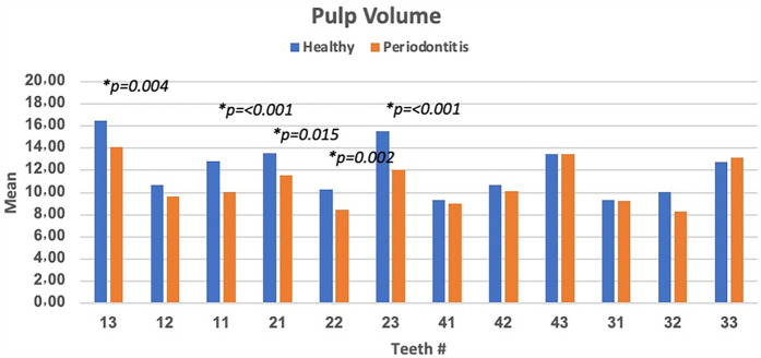
Results: The mean pulp volume between healthy and teeth with periodontitis showed certain differences. The mean low and largest pulp volumes of 9.15 ± 3.3 mm3 and 15.24 ± 4.2 mm3 were observed involving teeth # 41 and teeth # 13, respectively. Furthermore, a higher mean of pulp volume was observed in healthy teeth than in periodontitis-diagnosed teeth except for teeth # 33 and 43. The significant difference (p < 0.05) was easily detected involving teeth # 22, 23, 11, and 13. However, the lowest difference, with non-significant difference (p > 0.05), involving teeth # 43, 31, and 33 was observed.
Discussion: The study's findings underscore a significant correlation between periodontitis and reduced pulp volume, suggesting that periodontal inflammation may influence pupal remodeling.

 求助内容:
求助内容: 应助结果提醒方式:


