{"title":"有肉芽肿特征的纵隔精原细胞瘤1例:诊断和治疗的考虑。","authors":"Ryusei Yoshino, Nanami Ujiie, Shunsuke Yasuda, Yuki Kamikokura, Masahiro Kitada","doi":"10.70352/scrj.cr.25-0091","DOIUrl":null,"url":null,"abstract":"<p><strong>Introduction: </strong>Although seminomas typically arise in the testes, primary mediastinal seminomas are classified as extragonadal germ cell tumors. Diagnosis is often challenging and requires not only blood tests and imaging but also a tumor biopsy. However, diagnosis may be particularly difficult when the tumor shows nonspecific pathological features or is accompanied by granulomatous changes.</p><p><strong>Case presentation: </strong>The patient was a 25-year-old man who had been experiencing labored breathing when leaning forward for the past month. Physical examination revealed distended jugular veins and neck edema. Chest computed tomography revealed an irregular mass measuring 80 mm in the anterior mediastinum, suggesting invasion of the superior vena cava. Additionally, fluorodeoxyglucose-positron emission tomography showed high accumulation in the same area, with a maximum standardized uptake value of 11.3. A tumor biopsy was performed under thoracoscopic guidance for definitive diagnosis. Histopathological examination of the resected specimen revealed a seminoma with granulomatous changes. Based on these findings, a diagnosis of anterior mediastinal seminoma with superior vena cava syndrome was made. It was classified as having a good prognosis, and the patient received three courses of induction chemotherapy with etoposide, cisplatin, and ifosfamide. Complete remission was achieved. Since then, the patient has been monitored every 3 months, with no recurrence or metastasis observed for approximately 2 years.</p><p><strong>Conclusions: </strong>Immunohistochemical analysis plays a crucial role in the accurate diagnosis of mediastinal seminomas, especially in cases with unusual histological features such as granulomatous changes. Recognizing the immunoprofile of seminomas and differentiating them from thymomas and lymphomas is essential for avoiding diagnostic pitfalls.</p>","PeriodicalId":22096,"journal":{"name":"Surgical Case Reports","volume":"11 1","pages":""},"PeriodicalIF":0.7000,"publicationDate":"2025-01-01","publicationTypes":"Journal Article","fieldsOfStudy":null,"isOpenAccess":false,"openAccessPdf":"https://www.ncbi.nlm.nih.gov/pmc/articles/PMC12226142/pdf/","citationCount":"0","resultStr":"{\"title\":\"A Rare Case of Mediastinal Seminoma with Granulomatous Features: Diagnostic and Therapeutic Considerations.\",\"authors\":\"Ryusei Yoshino, Nanami Ujiie, Shunsuke Yasuda, Yuki Kamikokura, Masahiro Kitada\",\"doi\":\"10.70352/scrj.cr.25-0091\",\"DOIUrl\":null,\"url\":null,\"abstract\":\"<p><strong>Introduction: </strong>Although seminomas typically arise in the testes, primary mediastinal seminomas are classified as extragonadal germ cell tumors. Diagnosis is often challenging and requires not only blood tests and imaging but also a tumor biopsy. However, diagnosis may be particularly difficult when the tumor shows nonspecific pathological features or is accompanied by granulomatous changes.</p><p><strong>Case presentation: </strong>The patient was a 25-year-old man who had been experiencing labored breathing when leaning forward for the past month. Physical examination revealed distended jugular veins and neck edema. Chest computed tomography revealed an irregular mass measuring 80 mm in the anterior mediastinum, suggesting invasion of the superior vena cava. Additionally, fluorodeoxyglucose-positron emission tomography showed high accumulation in the same area, with a maximum standardized uptake value of 11.3. A tumor biopsy was performed under thoracoscopic guidance for definitive diagnosis. Histopathological examination of the resected specimen revealed a seminoma with granulomatous changes. Based on these findings, a diagnosis of anterior mediastinal seminoma with superior vena cava syndrome was made. It was classified as having a good prognosis, and the patient received three courses of induction chemotherapy with etoposide, cisplatin, and ifosfamide. Complete remission was achieved. Since then, the patient has been monitored every 3 months, with no recurrence or metastasis observed for approximately 2 years.</p><p><strong>Conclusions: </strong>Immunohistochemical analysis plays a crucial role in the accurate diagnosis of mediastinal seminomas, especially in cases with unusual histological features such as granulomatous changes. Recognizing the immunoprofile of seminomas and differentiating them from thymomas and lymphomas is essential for avoiding diagnostic pitfalls.</p>\",\"PeriodicalId\":22096,\"journal\":{\"name\":\"Surgical Case Reports\",\"volume\":\"11 1\",\"pages\":\"\"},\"PeriodicalIF\":0.7000,\"publicationDate\":\"2025-01-01\",\"publicationTypes\":\"Journal Article\",\"fieldsOfStudy\":null,\"isOpenAccess\":false,\"openAccessPdf\":\"https://www.ncbi.nlm.nih.gov/pmc/articles/PMC12226142/pdf/\",\"citationCount\":\"0\",\"resultStr\":null,\"platform\":\"Semanticscholar\",\"paperid\":null,\"PeriodicalName\":\"Surgical Case Reports\",\"FirstCategoryId\":\"1085\",\"ListUrlMain\":\"https://doi.org/10.70352/scrj.cr.25-0091\",\"RegionNum\":0,\"RegionCategory\":null,\"ArticlePicture\":[],\"TitleCN\":null,\"AbstractTextCN\":null,\"PMCID\":null,\"EPubDate\":\"2025/7/1 0:00:00\",\"PubModel\":\"Epub\",\"JCR\":\"Q4\",\"JCRName\":\"SURGERY\",\"Score\":null,\"Total\":0}","platform":"Semanticscholar","paperid":null,"PeriodicalName":"Surgical Case Reports","FirstCategoryId":"1085","ListUrlMain":"https://doi.org/10.70352/scrj.cr.25-0091","RegionNum":0,"RegionCategory":null,"ArticlePicture":[],"TitleCN":null,"AbstractTextCN":null,"PMCID":null,"EPubDate":"2025/7/1 0:00:00","PubModel":"Epub","JCR":"Q4","JCRName":"SURGERY","Score":null,"Total":0}
A Rare Case of Mediastinal Seminoma with Granulomatous Features: Diagnostic and Therapeutic Considerations.
Introduction: Although seminomas typically arise in the testes, primary mediastinal seminomas are classified as extragonadal germ cell tumors. Diagnosis is often challenging and requires not only blood tests and imaging but also a tumor biopsy. However, diagnosis may be particularly difficult when the tumor shows nonspecific pathological features or is accompanied by granulomatous changes.
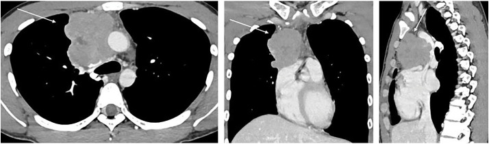
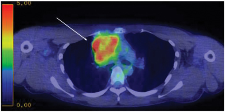
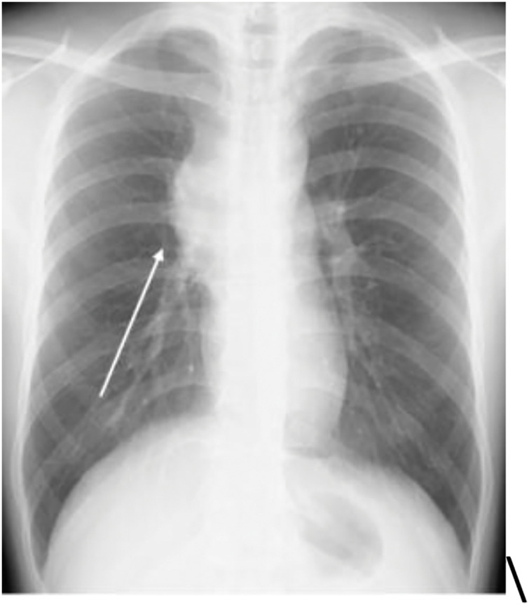
Case presentation: The patient was a 25-year-old man who had been experiencing labored breathing when leaning forward for the past month. Physical examination revealed distended jugular veins and neck edema. Chest computed tomography revealed an irregular mass measuring 80 mm in the anterior mediastinum, suggesting invasion of the superior vena cava. Additionally, fluorodeoxyglucose-positron emission tomography showed high accumulation in the same area, with a maximum standardized uptake value of 11.3. A tumor biopsy was performed under thoracoscopic guidance for definitive diagnosis. Histopathological examination of the resected specimen revealed a seminoma with granulomatous changes. Based on these findings, a diagnosis of anterior mediastinal seminoma with superior vena cava syndrome was made. It was classified as having a good prognosis, and the patient received three courses of induction chemotherapy with etoposide, cisplatin, and ifosfamide. Complete remission was achieved. Since then, the patient has been monitored every 3 months, with no recurrence or metastasis observed for approximately 2 years.
Conclusions: Immunohistochemical analysis plays a crucial role in the accurate diagnosis of mediastinal seminomas, especially in cases with unusual histological features such as granulomatous changes. Recognizing the immunoprofile of seminomas and differentiating them from thymomas and lymphomas is essential for avoiding diagnostic pitfalls.

 求助内容:
求助内容: 应助结果提醒方式:


