Chang Liu, Yang Sun, Yongyue Zhang, Rongjin Zhang, Liwei Li, Shumin Wang, Huiying He
{"title":"典型血管平滑肌脂肪瘤合并静脉肿瘤血栓形成的临床及影像学特征:与肾细胞癌的对比分析。","authors":"Chang Liu, Yang Sun, Yongyue Zhang, Rongjin Zhang, Liwei Li, Shumin Wang, Huiying He","doi":"10.1186/s12894-025-01833-4","DOIUrl":null,"url":null,"abstract":"<p><strong>Objective: </strong>To retrospectively analyze the clinical and imaging characteristics of classic angiomyolipoma (CAML) with venous tumor thrombus (VTT) and compare them with those of clear cell renal cell carcinoma (ccRCC).</p><p><strong>Methods: </strong>Clinical data from six patients with renal CAML complicated by VTT and 18 with ccRCC complicated by VTT, treated at Peking University Third Hospital from April 2018 and June 2022, were retrospectively analyzed. All patients underwent preoperative ultrasound and contrast-enhanced CT. Clinical manifestations, imaging characteristics, surgical findings, and pathological data were collected, and patients were followed up.</p><p><strong>Results: </strong>Enhanced CT showed renal sinus involvement in all CAML cases versus four ccRCC cases (p = 0.002). All primary CAML tumors had fatty components, compared to one ccRCC case (p < 0.001). Enhanced CT also revealed 7 VTTs with fatty components (6 in the CAML group) (p < 0.001). Thrombus lengths in the inferior vena cava (IVC) were 8.05 ± 2.22 cm for CAML and 5.29 ± 2.38 cm for ccRCC, with no significant difference (p = 0.610). The maximum/minimum anteroposterior VTT diameter ratios were 3.98 and 1.09, respectively (p < 0.001); coronal diameter ratios were 4.00 and 1.12, respectively (p < 0.001). Ultrasound revealed that, except for one Mayo Level 0 case, the involved IVC in the CAML group had continuous, intact walls with blood flow signals in the residual lumen, while in the ccRCC group, most VTTs had unclear boundaries and only one case showed blood flow signals in the residual lumen (p = 0.001). Intraoperative blood loss was significantly lower in CAML cases (p = 0.017). No CAML patient had VTT invading the venous wall, unlike 8 ccRCC patients (p = 0.016). All patients were followed for 21-74 months (median: 34.5 months, mean: 36.6 months). All were alive with normal renal function, and no tumor recurrence or metastasis was observed.</p><p><strong>Conclusion: </strong>Renal CAML with VTT is characterized by three imaging features: the presence of fatty components, a unique geometric growth pattern, and the absence of venous wall invasion, potentially serving as valuable indicators for differentiating CAML from ccRCC lesions.</p>","PeriodicalId":9285,"journal":{"name":"BMC Urology","volume":"25 1","pages":"151"},"PeriodicalIF":1.9000,"publicationDate":"2025-07-03","publicationTypes":"Journal Article","fieldsOfStudy":null,"isOpenAccess":false,"openAccessPdf":"https://www.ncbi.nlm.nih.gov/pmc/articles/PMC12224625/pdf/","citationCount":"0","resultStr":"{\"title\":\"Clinical and imaging characteristics of classic angiomyolipoma with venous tumor thrombosis: a comparative analysis against renal cell carcinoma.\",\"authors\":\"Chang Liu, Yang Sun, Yongyue Zhang, Rongjin Zhang, Liwei Li, Shumin Wang, Huiying He\",\"doi\":\"10.1186/s12894-025-01833-4\",\"DOIUrl\":null,\"url\":null,\"abstract\":\"<p><strong>Objective: </strong>To retrospectively analyze the clinical and imaging characteristics of classic angiomyolipoma (CAML) with venous tumor thrombus (VTT) and compare them with those of clear cell renal cell carcinoma (ccRCC).</p><p><strong>Methods: </strong>Clinical data from six patients with renal CAML complicated by VTT and 18 with ccRCC complicated by VTT, treated at Peking University Third Hospital from April 2018 and June 2022, were retrospectively analyzed. All patients underwent preoperative ultrasound and contrast-enhanced CT. Clinical manifestations, imaging characteristics, surgical findings, and pathological data were collected, and patients were followed up.</p><p><strong>Results: </strong>Enhanced CT showed renal sinus involvement in all CAML cases versus four ccRCC cases (p = 0.002). All primary CAML tumors had fatty components, compared to one ccRCC case (p < 0.001). Enhanced CT also revealed 7 VTTs with fatty components (6 in the CAML group) (p < 0.001). Thrombus lengths in the inferior vena cava (IVC) were 8.05 ± 2.22 cm for CAML and 5.29 ± 2.38 cm for ccRCC, with no significant difference (p = 0.610). The maximum/minimum anteroposterior VTT diameter ratios were 3.98 and 1.09, respectively (p < 0.001); coronal diameter ratios were 4.00 and 1.12, respectively (p < 0.001). Ultrasound revealed that, except for one Mayo Level 0 case, the involved IVC in the CAML group had continuous, intact walls with blood flow signals in the residual lumen, while in the ccRCC group, most VTTs had unclear boundaries and only one case showed blood flow signals in the residual lumen (p = 0.001). Intraoperative blood loss was significantly lower in CAML cases (p = 0.017). No CAML patient had VTT invading the venous wall, unlike 8 ccRCC patients (p = 0.016). All patients were followed for 21-74 months (median: 34.5 months, mean: 36.6 months). All were alive with normal renal function, and no tumor recurrence or metastasis was observed.</p><p><strong>Conclusion: </strong>Renal CAML with VTT is characterized by three imaging features: the presence of fatty components, a unique geometric growth pattern, and the absence of venous wall invasion, potentially serving as valuable indicators for differentiating CAML from ccRCC lesions.</p>\",\"PeriodicalId\":9285,\"journal\":{\"name\":\"BMC Urology\",\"volume\":\"25 1\",\"pages\":\"151\"},\"PeriodicalIF\":1.9000,\"publicationDate\":\"2025-07-03\",\"publicationTypes\":\"Journal Article\",\"fieldsOfStudy\":null,\"isOpenAccess\":false,\"openAccessPdf\":\"https://www.ncbi.nlm.nih.gov/pmc/articles/PMC12224625/pdf/\",\"citationCount\":\"0\",\"resultStr\":null,\"platform\":\"Semanticscholar\",\"paperid\":null,\"PeriodicalName\":\"BMC Urology\",\"FirstCategoryId\":\"3\",\"ListUrlMain\":\"https://doi.org/10.1186/s12894-025-01833-4\",\"RegionNum\":3,\"RegionCategory\":\"医学\",\"ArticlePicture\":[],\"TitleCN\":null,\"AbstractTextCN\":null,\"PMCID\":null,\"EPubDate\":\"\",\"PubModel\":\"\",\"JCR\":\"Q3\",\"JCRName\":\"UROLOGY & NEPHROLOGY\",\"Score\":null,\"Total\":0}","platform":"Semanticscholar","paperid":null,"PeriodicalName":"BMC Urology","FirstCategoryId":"3","ListUrlMain":"https://doi.org/10.1186/s12894-025-01833-4","RegionNum":3,"RegionCategory":"医学","ArticlePicture":[],"TitleCN":null,"AbstractTextCN":null,"PMCID":null,"EPubDate":"","PubModel":"","JCR":"Q3","JCRName":"UROLOGY & NEPHROLOGY","Score":null,"Total":0}
Clinical and imaging characteristics of classic angiomyolipoma with venous tumor thrombosis: a comparative analysis against renal cell carcinoma.
Objective: To retrospectively analyze the clinical and imaging characteristics of classic angiomyolipoma (CAML) with venous tumor thrombus (VTT) and compare them with those of clear cell renal cell carcinoma (ccRCC).
Methods: Clinical data from six patients with renal CAML complicated by VTT and 18 with ccRCC complicated by VTT, treated at Peking University Third Hospital from April 2018 and June 2022, were retrospectively analyzed. All patients underwent preoperative ultrasound and contrast-enhanced CT. Clinical manifestations, imaging characteristics, surgical findings, and pathological data were collected, and patients were followed up.
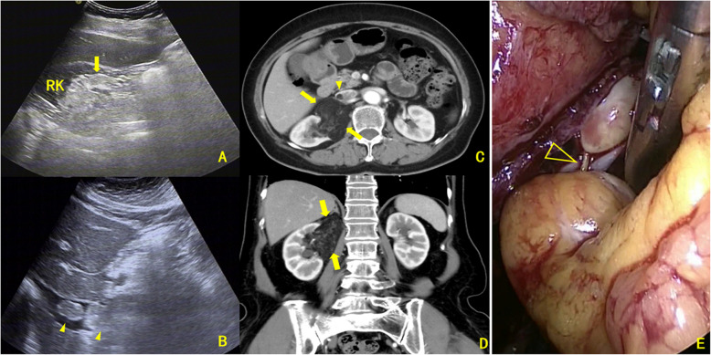
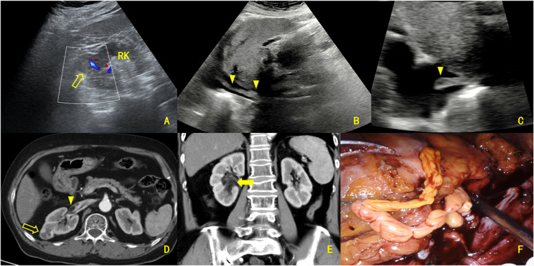
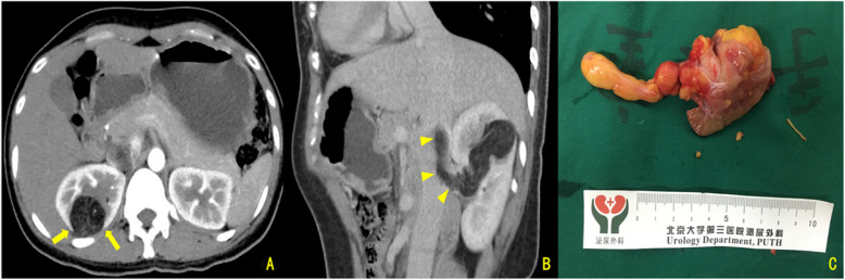
Results: Enhanced CT showed renal sinus involvement in all CAML cases versus four ccRCC cases (p = 0.002). All primary CAML tumors had fatty components, compared to one ccRCC case (p < 0.001). Enhanced CT also revealed 7 VTTs with fatty components (6 in the CAML group) (p < 0.001). Thrombus lengths in the inferior vena cava (IVC) were 8.05 ± 2.22 cm for CAML and 5.29 ± 2.38 cm for ccRCC, with no significant difference (p = 0.610). The maximum/minimum anteroposterior VTT diameter ratios were 3.98 and 1.09, respectively (p < 0.001); coronal diameter ratios were 4.00 and 1.12, respectively (p < 0.001). Ultrasound revealed that, except for one Mayo Level 0 case, the involved IVC in the CAML group had continuous, intact walls with blood flow signals in the residual lumen, while in the ccRCC group, most VTTs had unclear boundaries and only one case showed blood flow signals in the residual lumen (p = 0.001). Intraoperative blood loss was significantly lower in CAML cases (p = 0.017). No CAML patient had VTT invading the venous wall, unlike 8 ccRCC patients (p = 0.016). All patients were followed for 21-74 months (median: 34.5 months, mean: 36.6 months). All were alive with normal renal function, and no tumor recurrence or metastasis was observed.
Conclusion: Renal CAML with VTT is characterized by three imaging features: the presence of fatty components, a unique geometric growth pattern, and the absence of venous wall invasion, potentially serving as valuable indicators for differentiating CAML from ccRCC lesions.
期刊介绍:
BMC Urology is an open access journal publishing original peer-reviewed research articles in all aspects of the prevention, diagnosis and management of urological disorders, as well as related molecular genetics, pathophysiology, and epidemiology.
The journal considers manuscripts in the following broad subject-specific sections of urology:
Endourology and technology
Epidemiology and health outcomes
Pediatric urology
Pre-clinical and basic research
Reconstructive urology
Sexual function and fertility
Urological imaging
Urological oncology
Voiding dysfunction
Case reports.

 求助内容:
求助内容: 应助结果提醒方式:


