Samar F. Miski, Ahmed Serag, Arwa Sultan Alqahtani, Maram H. Abduljabbar, Reem M. Alnemari, Rami M. Alzhrani and Atiah H. Almalki
{"title":"关闭荧光感应雷洛昔芬使用红酶B与详细的光谱和量子力学研究的制药和环境应用†","authors":"Samar F. Miski, Ahmed Serag, Arwa Sultan Alqahtani, Maram H. Abduljabbar, Reem M. Alnemari, Rami M. Alzhrani and Atiah H. Almalki","doi":"10.1039/D5RA03551A","DOIUrl":null,"url":null,"abstract":"<p >A novel spectrofluorimetric method was developed for the detection of raloxifene based on its ability to quench the native fluorescence of the erythrosine B dye. Upon excitation at 528 nm, erythrosine B exhibits an emission peak at 554 nm, which undergoes concentration-dependent quenching upon interaction with raloxifene. Spectroscopic and thermodynamic studies revealed a static quenching mechanism with a Stern–Volmer constant of 4.87 × 10<small><sup>5</sup></small> M<small><sup>−1</sup></small> and a favorable Gibbs free energy change (Δ<em>G</em>) of −32.45 kJ mol<small><sup>−1</sup></small>. The calculated bimolecular quenching rate constant exceeded the diffusion-controlled limit, further confirming a ground-state complex formation. Job's method confirmed a 1 : 1 stoichiometric ratio, while quantum mechanical calculations elucidated the binding interactions with a binding energy of −0.143391 hartree and a reduction in dipole moment from 14.06 and 21.85 debye for erythrosine B and raloxifene, respectively, to 9.83 debye for the complex. Parameters affecting fluorescence quenching, such as pH, buffer volume, and erythrosine B concentration, were optimized, revealing maximum quenching at pH 4.0 using an acetate buffer, which is explained by the optimal ionization states of both molecules at this pH. The method validation as per ICH guidelines demonstrated linearity (0.1–3.0 μg mL<small><sup>−1</sup></small>, <em>r</em><small><sup>2</sup></small> = 0.9997), sensitivity (LOD = 0.0312 μg mL<small><sup>−1</sup></small>), accuracy (100.76% ± 1.277%), and precision (RSD < 1.671%). Analysis of pharmaceutical formulations showed 99.802% ± 0.528% recovery, with no significant difference from the reference HPLC method. The method was successfully applied to spiked plasma and environmental water samples with recoveries of 95.55–103.03% and 94.62–103.30%, respectively. AGREE calculator assessment (0.73) and BAGI (75.0) confirmed the greenness and practical applicability of the method, offering advantages of rapid analysis time (3 min) and minimal organic solvent consumption compared to existing techniques. This erythrosine B-based approach presents a viable alternative for raloxifene determination in resource-limited settings across diverse sample matrices.</p>","PeriodicalId":102,"journal":{"name":"RSC Advances","volume":" 29","pages":" 23124-23135"},"PeriodicalIF":4.6000,"publicationDate":"2025-07-04","publicationTypes":"Journal Article","fieldsOfStudy":null,"isOpenAccess":false,"openAccessPdf":"https://pubs.rsc.org/en/content/articlepdf/2025/ra/d5ra03551a?page=search","citationCount":"0","resultStr":"{\"title\":\"Turn-off fluorescence sensing of raloxifene using erythrosine B with detailed spectroscopic and quantum mechanical studies for pharmaceutical and environmental applications†\",\"authors\":\"Samar F. Miski, Ahmed Serag, Arwa Sultan Alqahtani, Maram H. Abduljabbar, Reem M. Alnemari, Rami M. Alzhrani and Atiah H. Almalki\",\"doi\":\"10.1039/D5RA03551A\",\"DOIUrl\":null,\"url\":null,\"abstract\":\"<p >A novel spectrofluorimetric method was developed for the detection of raloxifene based on its ability to quench the native fluorescence of the erythrosine B dye. Upon excitation at 528 nm, erythrosine B exhibits an emission peak at 554 nm, which undergoes concentration-dependent quenching upon interaction with raloxifene. Spectroscopic and thermodynamic studies revealed a static quenching mechanism with a Stern–Volmer constant of 4.87 × 10<small><sup>5</sup></small> M<small><sup>−1</sup></small> and a favorable Gibbs free energy change (Δ<em>G</em>) of −32.45 kJ mol<small><sup>−1</sup></small>. The calculated bimolecular quenching rate constant exceeded the diffusion-controlled limit, further confirming a ground-state complex formation. Job's method confirmed a 1 : 1 stoichiometric ratio, while quantum mechanical calculations elucidated the binding interactions with a binding energy of −0.143391 hartree and a reduction in dipole moment from 14.06 and 21.85 debye for erythrosine B and raloxifene, respectively, to 9.83 debye for the complex. Parameters affecting fluorescence quenching, such as pH, buffer volume, and erythrosine B concentration, were optimized, revealing maximum quenching at pH 4.0 using an acetate buffer, which is explained by the optimal ionization states of both molecules at this pH. The method validation as per ICH guidelines demonstrated linearity (0.1–3.0 μg mL<small><sup>−1</sup></small>, <em>r</em><small><sup>2</sup></small> = 0.9997), sensitivity (LOD = 0.0312 μg mL<small><sup>−1</sup></small>), accuracy (100.76% ± 1.277%), and precision (RSD < 1.671%). Analysis of pharmaceutical formulations showed 99.802% ± 0.528% recovery, with no significant difference from the reference HPLC method. The method was successfully applied to spiked plasma and environmental water samples with recoveries of 95.55–103.03% and 94.62–103.30%, respectively. AGREE calculator assessment (0.73) and BAGI (75.0) confirmed the greenness and practical applicability of the method, offering advantages of rapid analysis time (3 min) and minimal organic solvent consumption compared to existing techniques. This erythrosine B-based approach presents a viable alternative for raloxifene determination in resource-limited settings across diverse sample matrices.</p>\",\"PeriodicalId\":102,\"journal\":{\"name\":\"RSC Advances\",\"volume\":\" 29\",\"pages\":\" 23124-23135\"},\"PeriodicalIF\":4.6000,\"publicationDate\":\"2025-07-04\",\"publicationTypes\":\"Journal Article\",\"fieldsOfStudy\":null,\"isOpenAccess\":false,\"openAccessPdf\":\"https://pubs.rsc.org/en/content/articlepdf/2025/ra/d5ra03551a?page=search\",\"citationCount\":\"0\",\"resultStr\":null,\"platform\":\"Semanticscholar\",\"paperid\":null,\"PeriodicalName\":\"RSC Advances\",\"FirstCategoryId\":\"92\",\"ListUrlMain\":\"https://pubs.rsc.org/en/content/articlelanding/2025/ra/d5ra03551a\",\"RegionNum\":3,\"RegionCategory\":\"化学\",\"ArticlePicture\":[],\"TitleCN\":null,\"AbstractTextCN\":null,\"PMCID\":null,\"EPubDate\":\"\",\"PubModel\":\"\",\"JCR\":\"Q2\",\"JCRName\":\"CHEMISTRY, MULTIDISCIPLINARY\",\"Score\":null,\"Total\":0}","platform":"Semanticscholar","paperid":null,"PeriodicalName":"RSC Advances","FirstCategoryId":"92","ListUrlMain":"https://pubs.rsc.org/en/content/articlelanding/2025/ra/d5ra03551a","RegionNum":3,"RegionCategory":"化学","ArticlePicture":[],"TitleCN":null,"AbstractTextCN":null,"PMCID":null,"EPubDate":"","PubModel":"","JCR":"Q2","JCRName":"CHEMISTRY, MULTIDISCIPLINARY","Score":null,"Total":0}
Turn-off fluorescence sensing of raloxifene using erythrosine B with detailed spectroscopic and quantum mechanical studies for pharmaceutical and environmental applications†
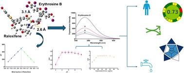
A novel spectrofluorimetric method was developed for the detection of raloxifene based on its ability to quench the native fluorescence of the erythrosine B dye. Upon excitation at 528 nm, erythrosine B exhibits an emission peak at 554 nm, which undergoes concentration-dependent quenching upon interaction with raloxifene. Spectroscopic and thermodynamic studies revealed a static quenching mechanism with a Stern–Volmer constant of 4.87 × 10⁵ M−1 and a favorable Gibbs free energy change (ΔG) of −32.45 kJ mol−1. The calculated bimolecular quenching rate constant exceeded the diffusion-controlled limit, further confirming a ground-state complex formation. Job's method confirmed a 1 : 1 stoichiometric ratio, while quantum mechanical calculations elucidated the binding interactions with a binding energy of −0.143391 hartree and a reduction in dipole moment from 14.06 and 21.85 debye for erythrosine B and raloxifene, respectively, to 9.83 debye for the complex. Parameters affecting fluorescence quenching, such as pH, buffer volume, and erythrosine B concentration, were optimized, revealing maximum quenching at pH 4.0 using an acetate buffer, which is explained by the optimal ionization states of both molecules at this pH. The method validation as per ICH guidelines demonstrated linearity (0.1–3.0 μg mL−1, r2 = 0.9997), sensitivity (LOD = 0.0312 μg mL−1), accuracy (100.76% ± 1.277%), and precision (RSD < 1.671%). Analysis of pharmaceutical formulations showed 99.802% ± 0.528% recovery, with no significant difference from the reference HPLC method. The method was successfully applied to spiked plasma and environmental water samples with recoveries of 95.55–103.03% and 94.62–103.30%, respectively. AGREE calculator assessment (0.73) and BAGI (75.0) confirmed the greenness and practical applicability of the method, offering advantages of rapid analysis time (3 min) and minimal organic solvent consumption compared to existing techniques. This erythrosine B-based approach presents a viable alternative for raloxifene determination in resource-limited settings across diverse sample matrices.
期刊介绍:
An international, peer-reviewed journal covering all of the chemical sciences, including multidisciplinary and emerging areas. RSC Advances is a gold open access journal allowing researchers free access to research articles, and offering an affordable open access publishing option for authors around the world.
 求助内容:
求助内容: 应助结果提醒方式:


