Carla Barros de Oliveira, Thaiza Gonçalves Rocha, Andrea Vaz Braga Pintor, Marcela Baraúna Magno, Aline Corrêa Abrahão, Lucianne Cople Maia, Mário José Romañach, Maria Augusta Visconti
{"title":"使用分形分析评估根管治疗后根尖周骨形成:系统回顾和荟萃分析。","authors":"Carla Barros de Oliveira, Thaiza Gonçalves Rocha, Andrea Vaz Braga Pintor, Marcela Baraúna Magno, Aline Corrêa Abrahão, Lucianne Cople Maia, Mário José Romañach, Maria Augusta Visconti","doi":"10.5624/isd.20240221","DOIUrl":null,"url":null,"abstract":"<p><strong>Purpose: </strong>This study aimed to review, evaluate, and synthesize existing evidence on the effectiveness of fractal analysis (FA) in assessing bone formation in periapical lesions following endodontic treatment.</p><p><strong>Materials and methods: </strong>Two reviewers systematically searched 6 electronic databases and gray literature. Studies were deemed eligible if they implemented the desired intervention and included a follow-up period of at least 12 months. Methodological quality was assessed using tools from the Joanna Briggs Institute. The meta-analysis calculated the mean difference (MD) in FA measurements of periapical lesion regions before and after endodontic treatment, with subgroup analyses based on bone and treatment type. The GRADE tool was employed to evaluate the certainty of the evidence.</p><p><strong>Results: </strong>Ten studies were included for qualitative synthesis and 8 in the meta-analysis. Overall, the mean fractal dimension (FD) increased following 12 months of endodontic treatment, with an MD of 0.223 (95% CI: 0.100-0.346; <i>P</i><0.001; I<sup>2</sup>=99%). Subgroup analyses revealed significantly increases in mean FD values for lesions in the maxilla (<i>P</i><0.01) and for the treatment subgroup (<i>P</i><0.01). However, the certainty of evidence was classified as very low.</p><p><strong>Conclusion: </strong>The observed increase in mean FD 12 months post-endodontic treatment across all included studies indicates bone formation in the periapical lesion regions.</p>","PeriodicalId":51714,"journal":{"name":"Imaging Science in Dentistry","volume":"55 2","pages":"126-138"},"PeriodicalIF":2.1000,"publicationDate":"2025-06-01","publicationTypes":"Journal Article","fieldsOfStudy":null,"isOpenAccess":false,"openAccessPdf":"https://www.ncbi.nlm.nih.gov/pmc/articles/PMC12210115/pdf/","citationCount":"0","resultStr":"{\"title\":\"Using fractal analysis to assess periapical bone formation after endodontic treatment: A systematic review and meta-analysis.\",\"authors\":\"Carla Barros de Oliveira, Thaiza Gonçalves Rocha, Andrea Vaz Braga Pintor, Marcela Baraúna Magno, Aline Corrêa Abrahão, Lucianne Cople Maia, Mário José Romañach, Maria Augusta Visconti\",\"doi\":\"10.5624/isd.20240221\",\"DOIUrl\":null,\"url\":null,\"abstract\":\"<p><strong>Purpose: </strong>This study aimed to review, evaluate, and synthesize existing evidence on the effectiveness of fractal analysis (FA) in assessing bone formation in periapical lesions following endodontic treatment.</p><p><strong>Materials and methods: </strong>Two reviewers systematically searched 6 electronic databases and gray literature. Studies were deemed eligible if they implemented the desired intervention and included a follow-up period of at least 12 months. Methodological quality was assessed using tools from the Joanna Briggs Institute. The meta-analysis calculated the mean difference (MD) in FA measurements of periapical lesion regions before and after endodontic treatment, with subgroup analyses based on bone and treatment type. The GRADE tool was employed to evaluate the certainty of the evidence.</p><p><strong>Results: </strong>Ten studies were included for qualitative synthesis and 8 in the meta-analysis. Overall, the mean fractal dimension (FD) increased following 12 months of endodontic treatment, with an MD of 0.223 (95% CI: 0.100-0.346; <i>P</i><0.001; I<sup>2</sup>=99%). Subgroup analyses revealed significantly increases in mean FD values for lesions in the maxilla (<i>P</i><0.01) and for the treatment subgroup (<i>P</i><0.01). However, the certainty of evidence was classified as very low.</p><p><strong>Conclusion: </strong>The observed increase in mean FD 12 months post-endodontic treatment across all included studies indicates bone formation in the periapical lesion regions.</p>\",\"PeriodicalId\":51714,\"journal\":{\"name\":\"Imaging Science in Dentistry\",\"volume\":\"55 2\",\"pages\":\"126-138\"},\"PeriodicalIF\":2.1000,\"publicationDate\":\"2025-06-01\",\"publicationTypes\":\"Journal Article\",\"fieldsOfStudy\":null,\"isOpenAccess\":false,\"openAccessPdf\":\"https://www.ncbi.nlm.nih.gov/pmc/articles/PMC12210115/pdf/\",\"citationCount\":\"0\",\"resultStr\":null,\"platform\":\"Semanticscholar\",\"paperid\":null,\"PeriodicalName\":\"Imaging Science in Dentistry\",\"FirstCategoryId\":\"1085\",\"ListUrlMain\":\"https://doi.org/10.5624/isd.20240221\",\"RegionNum\":0,\"RegionCategory\":null,\"ArticlePicture\":[],\"TitleCN\":null,\"AbstractTextCN\":null,\"PMCID\":null,\"EPubDate\":\"2025/4/10 0:00:00\",\"PubModel\":\"Epub\",\"JCR\":\"Q3\",\"JCRName\":\"DENTISTRY, ORAL SURGERY & MEDICINE\",\"Score\":null,\"Total\":0}","platform":"Semanticscholar","paperid":null,"PeriodicalName":"Imaging Science in Dentistry","FirstCategoryId":"1085","ListUrlMain":"https://doi.org/10.5624/isd.20240221","RegionNum":0,"RegionCategory":null,"ArticlePicture":[],"TitleCN":null,"AbstractTextCN":null,"PMCID":null,"EPubDate":"2025/4/10 0:00:00","PubModel":"Epub","JCR":"Q3","JCRName":"DENTISTRY, ORAL SURGERY & MEDICINE","Score":null,"Total":0}
Using fractal analysis to assess periapical bone formation after endodontic treatment: A systematic review and meta-analysis.
Purpose: This study aimed to review, evaluate, and synthesize existing evidence on the effectiveness of fractal analysis (FA) in assessing bone formation in periapical lesions following endodontic treatment.
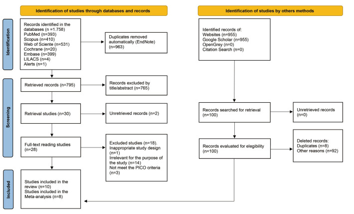
Materials and methods: Two reviewers systematically searched 6 electronic databases and gray literature. Studies were deemed eligible if they implemented the desired intervention and included a follow-up period of at least 12 months. Methodological quality was assessed using tools from the Joanna Briggs Institute. The meta-analysis calculated the mean difference (MD) in FA measurements of periapical lesion regions before and after endodontic treatment, with subgroup analyses based on bone and treatment type. The GRADE tool was employed to evaluate the certainty of the evidence.
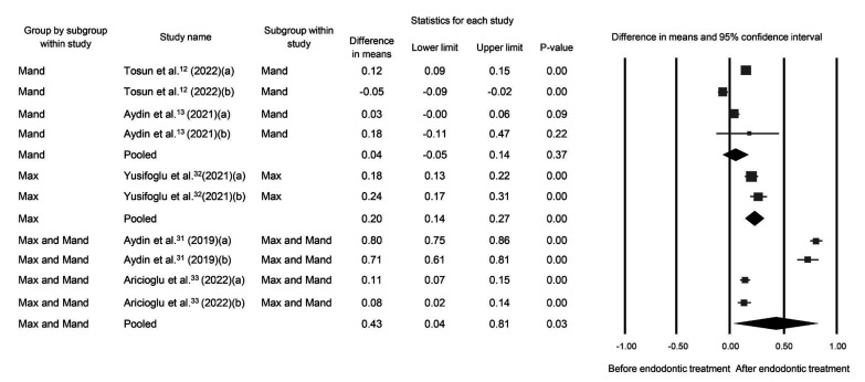
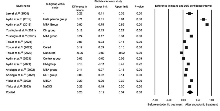
Results: Ten studies were included for qualitative synthesis and 8 in the meta-analysis. Overall, the mean fractal dimension (FD) increased following 12 months of endodontic treatment, with an MD of 0.223 (95% CI: 0.100-0.346; P<0.001; I2=99%). Subgroup analyses revealed significantly increases in mean FD values for lesions in the maxilla (P<0.01) and for the treatment subgroup (P<0.01). However, the certainty of evidence was classified as very low.
Conclusion: The observed increase in mean FD 12 months post-endodontic treatment across all included studies indicates bone formation in the periapical lesion regions.

 求助内容:
求助内容: 应助结果提醒方式:


