Medhana Mangaonker, Shamli Prabhu Chodnekar, Manisha M Khorate, Nigel Figueiredo, Miyola Cia Fernandes, Sushmita Wayadande
{"title":"磁共振弥散加权成像鉴别单囊性成釉细胞瘤、牙源性角化囊肿和牙源性囊肿:系统综述和网络荟萃分析。","authors":"Medhana Mangaonker, Shamli Prabhu Chodnekar, Manisha M Khorate, Nigel Figueiredo, Miyola Cia Fernandes, Sushmita Wayadande","doi":"10.5624/isd.20240227","DOIUrl":null,"url":null,"abstract":"<p><strong>Purpose: </strong>Diffusion-weighted magnetic resonance imaging (DW-MRI) facilitates the differentiation of unicystic ameloblastoma (UAM), odontogenic keratocyst (OKC), and dentigerous cyst (DC) by depicting detailed internal lesion structures based on water molecule movement. This study aimed to evaluate the efficacy of DW-MRI in distinguishing UAM, OKC, and DC.</p><p><strong>Materials and methods: </strong>This systematic review included studies from 2008 to 2022 that evaluated the diagnostic accuracy of DW-MRI through apparent diffusion coefficient (ADC) values in UAM, OKC, and DC. Six studies were qualitatively appraised using the QUADAS-2 tool, and 4 studies were subsequently included in a network meta-analysis for quantitative assessment of mean ADC values. The protocol was registered with PROSPERO (registration number: CRD42024502152).</p><p><strong>Results: </strong>Six studies encompassing 230 patients employed DW-MRI with an echo planar imaging sequence, yielding images with either hyperintense or hypointense lesion enhancements. The studies demonstrated that the mean ADC value for UAM was >2.0×10<sup>-3</sup> mm<sup>2</sup>/s, for DC was >1.0×10<sup>-3</sup> mm<sup>2</sup>/s, and for OKC was <1.0×10<sup>-3</sup> mm<sup>2</sup>/s (<i>P</i><0.05).</p><p><strong>Conclusion: </strong>This systematic review shows that DW-MRI, when used in conjunction with ADC measurements, effectively differentiates among UAM, OKC, and DC. The statistically significant ADC cut-off values support the use of DW-MRI as an adjunctive imaging modality to improve diagnostic accuracy in clinical practice.</p>","PeriodicalId":51714,"journal":{"name":"Imaging Science in Dentistry","volume":"55 2","pages":"105-113"},"PeriodicalIF":2.1000,"publicationDate":"2025-06-01","publicationTypes":"Journal Article","fieldsOfStudy":null,"isOpenAccess":false,"openAccessPdf":"https://www.ncbi.nlm.nih.gov/pmc/articles/PMC12210118/pdf/","citationCount":"0","resultStr":"{\"title\":\"Diffusion-weighted magnetic resonance imaging for differentiation of unicystic ameloblastoma, odontogenic keratocyst, and dentigerous cyst: A systematic review and network meta-analysis.\",\"authors\":\"Medhana Mangaonker, Shamli Prabhu Chodnekar, Manisha M Khorate, Nigel Figueiredo, Miyola Cia Fernandes, Sushmita Wayadande\",\"doi\":\"10.5624/isd.20240227\",\"DOIUrl\":null,\"url\":null,\"abstract\":\"<p><strong>Purpose: </strong>Diffusion-weighted magnetic resonance imaging (DW-MRI) facilitates the differentiation of unicystic ameloblastoma (UAM), odontogenic keratocyst (OKC), and dentigerous cyst (DC) by depicting detailed internal lesion structures based on water molecule movement. This study aimed to evaluate the efficacy of DW-MRI in distinguishing UAM, OKC, and DC.</p><p><strong>Materials and methods: </strong>This systematic review included studies from 2008 to 2022 that evaluated the diagnostic accuracy of DW-MRI through apparent diffusion coefficient (ADC) values in UAM, OKC, and DC. Six studies were qualitatively appraised using the QUADAS-2 tool, and 4 studies were subsequently included in a network meta-analysis for quantitative assessment of mean ADC values. The protocol was registered with PROSPERO (registration number: CRD42024502152).</p><p><strong>Results: </strong>Six studies encompassing 230 patients employed DW-MRI with an echo planar imaging sequence, yielding images with either hyperintense or hypointense lesion enhancements. The studies demonstrated that the mean ADC value for UAM was >2.0×10<sup>-3</sup> mm<sup>2</sup>/s, for DC was >1.0×10<sup>-3</sup> mm<sup>2</sup>/s, and for OKC was <1.0×10<sup>-3</sup> mm<sup>2</sup>/s (<i>P</i><0.05).</p><p><strong>Conclusion: </strong>This systematic review shows that DW-MRI, when used in conjunction with ADC measurements, effectively differentiates among UAM, OKC, and DC. The statistically significant ADC cut-off values support the use of DW-MRI as an adjunctive imaging modality to improve diagnostic accuracy in clinical practice.</p>\",\"PeriodicalId\":51714,\"journal\":{\"name\":\"Imaging Science in Dentistry\",\"volume\":\"55 2\",\"pages\":\"105-113\"},\"PeriodicalIF\":2.1000,\"publicationDate\":\"2025-06-01\",\"publicationTypes\":\"Journal Article\",\"fieldsOfStudy\":null,\"isOpenAccess\":false,\"openAccessPdf\":\"https://www.ncbi.nlm.nih.gov/pmc/articles/PMC12210118/pdf/\",\"citationCount\":\"0\",\"resultStr\":null,\"platform\":\"Semanticscholar\",\"paperid\":null,\"PeriodicalName\":\"Imaging Science in Dentistry\",\"FirstCategoryId\":\"1085\",\"ListUrlMain\":\"https://doi.org/10.5624/isd.20240227\",\"RegionNum\":0,\"RegionCategory\":null,\"ArticlePicture\":[],\"TitleCN\":null,\"AbstractTextCN\":null,\"PMCID\":null,\"EPubDate\":\"2025/4/28 0:00:00\",\"PubModel\":\"Epub\",\"JCR\":\"Q3\",\"JCRName\":\"DENTISTRY, ORAL SURGERY & MEDICINE\",\"Score\":null,\"Total\":0}","platform":"Semanticscholar","paperid":null,"PeriodicalName":"Imaging Science in Dentistry","FirstCategoryId":"1085","ListUrlMain":"https://doi.org/10.5624/isd.20240227","RegionNum":0,"RegionCategory":null,"ArticlePicture":[],"TitleCN":null,"AbstractTextCN":null,"PMCID":null,"EPubDate":"2025/4/28 0:00:00","PubModel":"Epub","JCR":"Q3","JCRName":"DENTISTRY, ORAL SURGERY & MEDICINE","Score":null,"Total":0}
Diffusion-weighted magnetic resonance imaging for differentiation of unicystic ameloblastoma, odontogenic keratocyst, and dentigerous cyst: A systematic review and network meta-analysis.
Purpose: Diffusion-weighted magnetic resonance imaging (DW-MRI) facilitates the differentiation of unicystic ameloblastoma (UAM), odontogenic keratocyst (OKC), and dentigerous cyst (DC) by depicting detailed internal lesion structures based on water molecule movement. This study aimed to evaluate the efficacy of DW-MRI in distinguishing UAM, OKC, and DC.
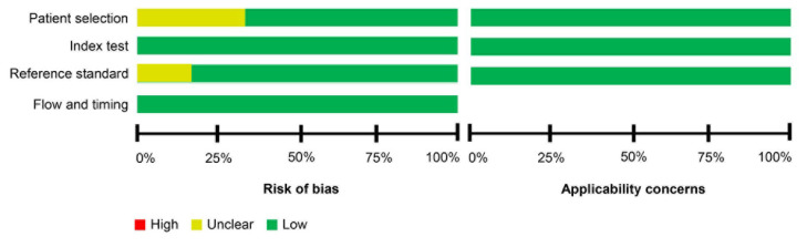
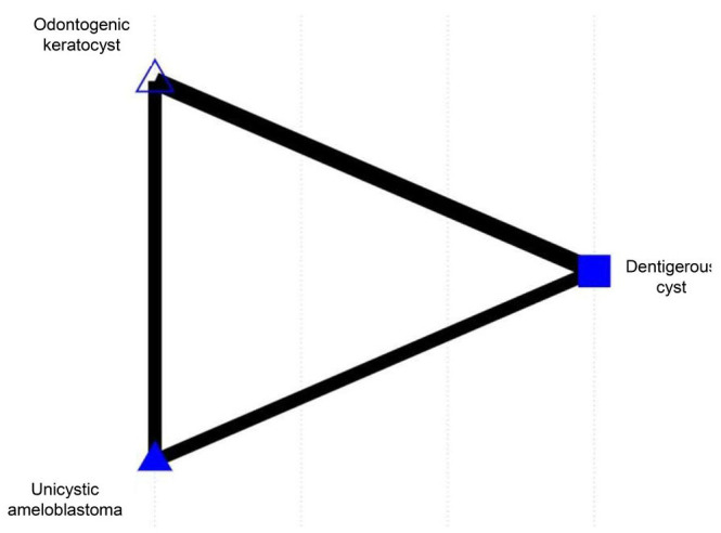
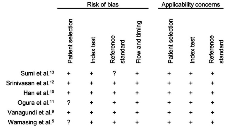
Materials and methods: This systematic review included studies from 2008 to 2022 that evaluated the diagnostic accuracy of DW-MRI through apparent diffusion coefficient (ADC) values in UAM, OKC, and DC. Six studies were qualitatively appraised using the QUADAS-2 tool, and 4 studies were subsequently included in a network meta-analysis for quantitative assessment of mean ADC values. The protocol was registered with PROSPERO (registration number: CRD42024502152).
Results: Six studies encompassing 230 patients employed DW-MRI with an echo planar imaging sequence, yielding images with either hyperintense or hypointense lesion enhancements. The studies demonstrated that the mean ADC value for UAM was >2.0×10⁻³ mm2/s, for DC was >1.0×10⁻³ mm2/s, and for OKC was <1.0×10⁻³ mm2/s (P<0.05).
Conclusion: This systematic review shows that DW-MRI, when used in conjunction with ADC measurements, effectively differentiates among UAM, OKC, and DC. The statistically significant ADC cut-off values support the use of DW-MRI as an adjunctive imaging modality to improve diagnostic accuracy in clinical practice.

 求助内容:
求助内容: 应助结果提醒方式:


