Matheus Sampaio-Oliveira, Matheus L Oliveira, Rubens Spin-Neto
{"title":"口腔专用磁共振成像载体装置的研制。","authors":"Matheus Sampaio-Oliveira, Matheus L Oliveira, Rubens Spin-Neto","doi":"10.5624/isd.20240254","DOIUrl":null,"url":null,"abstract":"<p><strong>Purpose: </strong>The aim of this study was to develop and evaluate a carrier device for dental-dedicated magnetic resonance imaging (ddMRI).</p><p><strong>Materials and methods: </strong>The carrier device comprised 5 glass test tubes, which were vertically positioned within a glass beaker and filled with air, distilled water, 1.5% agar, nickel nitrate [Ni(NO<sub>3</sub>)<sub>2</sub>] in 1.5% agar, or 1000 g·L<sup>-1</sup> dipotassium phosphate (K<sub>2</sub>HPO<sub>4</sub>). The beaker was filled with distilled water, a 0.3 g·L<sup>-1</sup> Ni(NO<sub>3</sub>)<sub>2</sub> aqueous solution, or a 1000 g·L<sup>-1</sup> K<sub>2</sub>HPO<sub>4</sub> aqueous solution. The device was scanned using a proton density turbo-spin-echo pulse sequence on a ddMRI system equipped with a dental-dedicated radiofrequency surface coil. Triplicate scans were performed for each combination of tube fillings and beaker solutions, yielding a total of 45 image volumes. Quantitative image metrics were then assessed.</p><p><strong>Results: </strong>The developed carrier device, composed of carrier vials filled with 1.5% agar surrounded by a 1000 g·L<sup>-1</sup> K<sub>2</sub>HPO<sub>4</sub> aqueous solution, was identified as the best option for ddMRI quality assessments.</p><p><strong>Conclusion: </strong>The proposed carrier device represents a promising method for embedding dental materials and other specimens, thereby facilitating the evaluation of their behaviour in ddMRI.</p>","PeriodicalId":51714,"journal":{"name":"Imaging Science in Dentistry","volume":"55 2","pages":"207-213"},"PeriodicalIF":2.1000,"publicationDate":"2025-06-01","publicationTypes":"Journal Article","fieldsOfStudy":null,"isOpenAccess":false,"openAccessPdf":"https://www.ncbi.nlm.nih.gov/pmc/articles/PMC12210122/pdf/","citationCount":"0","resultStr":"{\"title\":\"Development of a carrier device for dental-dedicated magnetic resonance imaging.\",\"authors\":\"Matheus Sampaio-Oliveira, Matheus L Oliveira, Rubens Spin-Neto\",\"doi\":\"10.5624/isd.20240254\",\"DOIUrl\":null,\"url\":null,\"abstract\":\"<p><strong>Purpose: </strong>The aim of this study was to develop and evaluate a carrier device for dental-dedicated magnetic resonance imaging (ddMRI).</p><p><strong>Materials and methods: </strong>The carrier device comprised 5 glass test tubes, which were vertically positioned within a glass beaker and filled with air, distilled water, 1.5% agar, nickel nitrate [Ni(NO<sub>3</sub>)<sub>2</sub>] in 1.5% agar, or 1000 g·L<sup>-1</sup> dipotassium phosphate (K<sub>2</sub>HPO<sub>4</sub>). The beaker was filled with distilled water, a 0.3 g·L<sup>-1</sup> Ni(NO<sub>3</sub>)<sub>2</sub> aqueous solution, or a 1000 g·L<sup>-1</sup> K<sub>2</sub>HPO<sub>4</sub> aqueous solution. The device was scanned using a proton density turbo-spin-echo pulse sequence on a ddMRI system equipped with a dental-dedicated radiofrequency surface coil. Triplicate scans were performed for each combination of tube fillings and beaker solutions, yielding a total of 45 image volumes. Quantitative image metrics were then assessed.</p><p><strong>Results: </strong>The developed carrier device, composed of carrier vials filled with 1.5% agar surrounded by a 1000 g·L<sup>-1</sup> K<sub>2</sub>HPO<sub>4</sub> aqueous solution, was identified as the best option for ddMRI quality assessments.</p><p><strong>Conclusion: </strong>The proposed carrier device represents a promising method for embedding dental materials and other specimens, thereby facilitating the evaluation of their behaviour in ddMRI.</p>\",\"PeriodicalId\":51714,\"journal\":{\"name\":\"Imaging Science in Dentistry\",\"volume\":\"55 2\",\"pages\":\"207-213\"},\"PeriodicalIF\":2.1000,\"publicationDate\":\"2025-06-01\",\"publicationTypes\":\"Journal Article\",\"fieldsOfStudy\":null,\"isOpenAccess\":false,\"openAccessPdf\":\"https://www.ncbi.nlm.nih.gov/pmc/articles/PMC12210122/pdf/\",\"citationCount\":\"0\",\"resultStr\":null,\"platform\":\"Semanticscholar\",\"paperid\":null,\"PeriodicalName\":\"Imaging Science in Dentistry\",\"FirstCategoryId\":\"1085\",\"ListUrlMain\":\"https://doi.org/10.5624/isd.20240254\",\"RegionNum\":0,\"RegionCategory\":null,\"ArticlePicture\":[],\"TitleCN\":null,\"AbstractTextCN\":null,\"PMCID\":null,\"EPubDate\":\"2025/4/10 0:00:00\",\"PubModel\":\"Epub\",\"JCR\":\"Q3\",\"JCRName\":\"DENTISTRY, ORAL SURGERY & MEDICINE\",\"Score\":null,\"Total\":0}","platform":"Semanticscholar","paperid":null,"PeriodicalName":"Imaging Science in Dentistry","FirstCategoryId":"1085","ListUrlMain":"https://doi.org/10.5624/isd.20240254","RegionNum":0,"RegionCategory":null,"ArticlePicture":[],"TitleCN":null,"AbstractTextCN":null,"PMCID":null,"EPubDate":"2025/4/10 0:00:00","PubModel":"Epub","JCR":"Q3","JCRName":"DENTISTRY, ORAL SURGERY & MEDICINE","Score":null,"Total":0}
Development of a carrier device for dental-dedicated magnetic resonance imaging.
Purpose: The aim of this study was to develop and evaluate a carrier device for dental-dedicated magnetic resonance imaging (ddMRI).
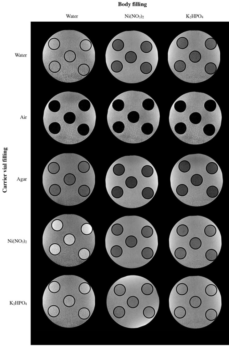
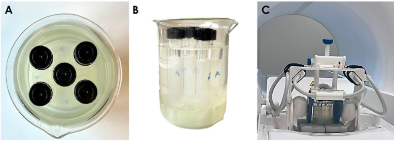
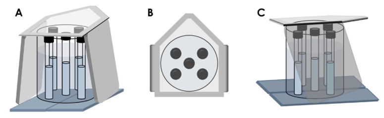
Materials and methods: The carrier device comprised 5 glass test tubes, which were vertically positioned within a glass beaker and filled with air, distilled water, 1.5% agar, nickel nitrate [Ni(NO3)2] in 1.5% agar, or 1000 g·L-1 dipotassium phosphate (K2HPO4). The beaker was filled with distilled water, a 0.3 g·L-1 Ni(NO3)2 aqueous solution, or a 1000 g·L-1 K2HPO4 aqueous solution. The device was scanned using a proton density turbo-spin-echo pulse sequence on a ddMRI system equipped with a dental-dedicated radiofrequency surface coil. Triplicate scans were performed for each combination of tube fillings and beaker solutions, yielding a total of 45 image volumes. Quantitative image metrics were then assessed.
Results: The developed carrier device, composed of carrier vials filled with 1.5% agar surrounded by a 1000 g·L-1 K2HPO4 aqueous solution, was identified as the best option for ddMRI quality assessments.
Conclusion: The proposed carrier device represents a promising method for embedding dental materials and other specimens, thereby facilitating the evaluation of their behaviour in ddMRI.

 求助内容:
求助内容: 应助结果提醒方式:


