{"title":"使用深度学习的牙科全景x光片自动质量评估。","authors":"Nazila Ameli, Masoud Miri Moghaddam, Hollis Lai, Camila Pacheco-Pereira","doi":"10.5624/isd.20240232","DOIUrl":null,"url":null,"abstract":"<p><strong>Purpose: </strong>Panoramic radiographs are instrumental in dental diagnosis but face quality issues related to contrast, artifacts, positioning, and coverage, which can impact diagnostic accuracy. Although expert assessment is the accepted standard, it is time-consuming and prone to inconsistency. Artificial intelligence offers an automated, objective solution for evaluating radiograph quality, increasing efficiency and reducing inter-rater variability.</p><p><strong>Materials and methods: </strong>This study aimed to develop a deep learning (DL)-based model for evaluating the quality of dental panoramic radiographs. A dataset of 1,000 panoramic images, collected from 2018 to 2023, was assessed by 2 trained dentists using predefined grading criteria for contrast/density, artifact presence, coverage area, patient positioning, and overall quality. These expert-annotated scores were used as the ground truth to train and validate 5 YOLOv8 classification models, each targeting a specific quality criterion. The models' performance was evaluated on a separate test set using performance metrics.</p><p><strong>Results: </strong>The YOLOv8 models achieved classification accuracies of 87.2%, 74.1%, 77.3%, 97.9%, and 79.3% for artifact detection, coverage area, patient positioning, contrast/density, and overall image quality, respectively. The model used to classify images as clinically acceptable or unacceptable exhibited an average accuracy of 81.4%, demonstrating its potential for real-world application.</p><p><strong>Conclusion: </strong>These findings highlight the feasibility of DL-based automated image quality assessment for panoramic radiographs. The high accuracy of the proposed model suggests its potential integration into clinical workflows to assist practitioners in efficiently evaluating radiograph quality. Additionally, such a model could represent an educational tool for dental students, improving radiographic techniques and reducing unnecessary retakes.</p>","PeriodicalId":51714,"journal":{"name":"Imaging Science in Dentistry","volume":"55 2","pages":"175-188"},"PeriodicalIF":2.1000,"publicationDate":"2025-06-01","publicationTypes":"Journal Article","fieldsOfStudy":null,"isOpenAccess":false,"openAccessPdf":"https://www.ncbi.nlm.nih.gov/pmc/articles/PMC12210116/pdf/","citationCount":"0","resultStr":"{\"title\":\"Automated quality evaluation of dental panoramic radiographs using deep learning.\",\"authors\":\"Nazila Ameli, Masoud Miri Moghaddam, Hollis Lai, Camila Pacheco-Pereira\",\"doi\":\"10.5624/isd.20240232\",\"DOIUrl\":null,\"url\":null,\"abstract\":\"<p><strong>Purpose: </strong>Panoramic radiographs are instrumental in dental diagnosis but face quality issues related to contrast, artifacts, positioning, and coverage, which can impact diagnostic accuracy. Although expert assessment is the accepted standard, it is time-consuming and prone to inconsistency. Artificial intelligence offers an automated, objective solution for evaluating radiograph quality, increasing efficiency and reducing inter-rater variability.</p><p><strong>Materials and methods: </strong>This study aimed to develop a deep learning (DL)-based model for evaluating the quality of dental panoramic radiographs. A dataset of 1,000 panoramic images, collected from 2018 to 2023, was assessed by 2 trained dentists using predefined grading criteria for contrast/density, artifact presence, coverage area, patient positioning, and overall quality. These expert-annotated scores were used as the ground truth to train and validate 5 YOLOv8 classification models, each targeting a specific quality criterion. The models' performance was evaluated on a separate test set using performance metrics.</p><p><strong>Results: </strong>The YOLOv8 models achieved classification accuracies of 87.2%, 74.1%, 77.3%, 97.9%, and 79.3% for artifact detection, coverage area, patient positioning, contrast/density, and overall image quality, respectively. The model used to classify images as clinically acceptable or unacceptable exhibited an average accuracy of 81.4%, demonstrating its potential for real-world application.</p><p><strong>Conclusion: </strong>These findings highlight the feasibility of DL-based automated image quality assessment for panoramic radiographs. The high accuracy of the proposed model suggests its potential integration into clinical workflows to assist practitioners in efficiently evaluating radiograph quality. Additionally, such a model could represent an educational tool for dental students, improving radiographic techniques and reducing unnecessary retakes.</p>\",\"PeriodicalId\":51714,\"journal\":{\"name\":\"Imaging Science in Dentistry\",\"volume\":\"55 2\",\"pages\":\"175-188\"},\"PeriodicalIF\":2.1000,\"publicationDate\":\"2025-06-01\",\"publicationTypes\":\"Journal Article\",\"fieldsOfStudy\":null,\"isOpenAccess\":false,\"openAccessPdf\":\"https://www.ncbi.nlm.nih.gov/pmc/articles/PMC12210116/pdf/\",\"citationCount\":\"0\",\"resultStr\":null,\"platform\":\"Semanticscholar\",\"paperid\":null,\"PeriodicalName\":\"Imaging Science in Dentistry\",\"FirstCategoryId\":\"1085\",\"ListUrlMain\":\"https://doi.org/10.5624/isd.20240232\",\"RegionNum\":0,\"RegionCategory\":null,\"ArticlePicture\":[],\"TitleCN\":null,\"AbstractTextCN\":null,\"PMCID\":null,\"EPubDate\":\"2025/4/10 0:00:00\",\"PubModel\":\"Epub\",\"JCR\":\"Q3\",\"JCRName\":\"DENTISTRY, ORAL SURGERY & MEDICINE\",\"Score\":null,\"Total\":0}","platform":"Semanticscholar","paperid":null,"PeriodicalName":"Imaging Science in Dentistry","FirstCategoryId":"1085","ListUrlMain":"https://doi.org/10.5624/isd.20240232","RegionNum":0,"RegionCategory":null,"ArticlePicture":[],"TitleCN":null,"AbstractTextCN":null,"PMCID":null,"EPubDate":"2025/4/10 0:00:00","PubModel":"Epub","JCR":"Q3","JCRName":"DENTISTRY, ORAL SURGERY & MEDICINE","Score":null,"Total":0}
Automated quality evaluation of dental panoramic radiographs using deep learning.
Purpose: Panoramic radiographs are instrumental in dental diagnosis but face quality issues related to contrast, artifacts, positioning, and coverage, which can impact diagnostic accuracy. Although expert assessment is the accepted standard, it is time-consuming and prone to inconsistency. Artificial intelligence offers an automated, objective solution for evaluating radiograph quality, increasing efficiency and reducing inter-rater variability.
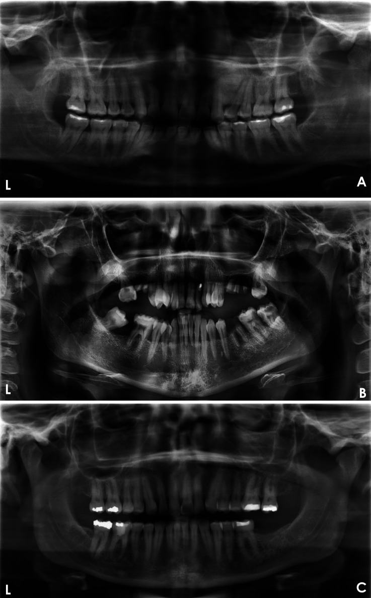
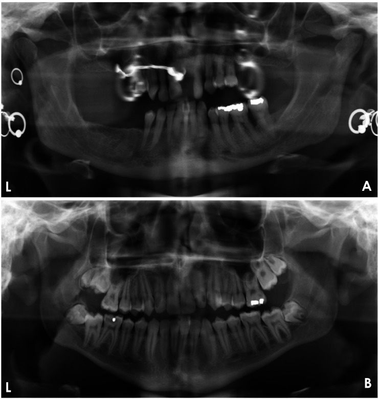
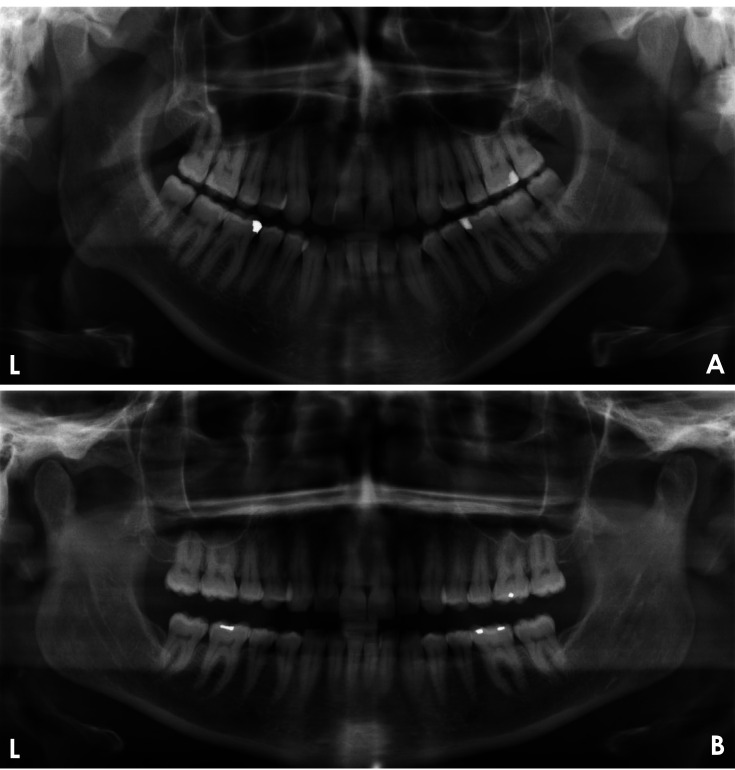
Materials and methods: This study aimed to develop a deep learning (DL)-based model for evaluating the quality of dental panoramic radiographs. A dataset of 1,000 panoramic images, collected from 2018 to 2023, was assessed by 2 trained dentists using predefined grading criteria for contrast/density, artifact presence, coverage area, patient positioning, and overall quality. These expert-annotated scores were used as the ground truth to train and validate 5 YOLOv8 classification models, each targeting a specific quality criterion. The models' performance was evaluated on a separate test set using performance metrics.
Results: The YOLOv8 models achieved classification accuracies of 87.2%, 74.1%, 77.3%, 97.9%, and 79.3% for artifact detection, coverage area, patient positioning, contrast/density, and overall image quality, respectively. The model used to classify images as clinically acceptable or unacceptable exhibited an average accuracy of 81.4%, demonstrating its potential for real-world application.
Conclusion: These findings highlight the feasibility of DL-based automated image quality assessment for panoramic radiographs. The high accuracy of the proposed model suggests its potential integration into clinical workflows to assist practitioners in efficiently evaluating radiograph quality. Additionally, such a model could represent an educational tool for dental students, improving radiographic techniques and reducing unnecessary retakes.

 求助内容:
求助内容: 应助结果提醒方式:


