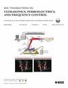
Hui Zhu, Yi Zeng, Jianfeng Li, Kailiang Xu, Xiran Cai
{"title":"基于交叉谱法的行-列阵列实时三维被动声映射。","authors":"Hui Zhu, Yi Zeng, Jianfeng Li, Kailiang Xu, Xiran Cai","doi":"10.1109/TUFFC.2025.3585301","DOIUrl":null,"url":null,"abstract":"<p><p>Real-time and three-dimensional (3D) monitoring of cavitation activity is critical for safe, effective and controlled treatments in cavitation-based focused ultrasound (FUS) therapies. This 3D monitoring capability is essential for detecting off-target cavitation events, particularly in at-risk structures and those occurring outside the plane of 2D imaging. In this work, we demonstrate that using row-column arrays (RCAs) for 3D passive acoustic mapping (PAM), which can be easily integrated to commercial ultrasound scanners compared to using hemispherical arrays or matrix arrays, represents a potent solution. For that, we propose the RCA-PAM method for image formation. This method deploys the angular spectrum (AS) method to back-propagate 3D harmonic wave fields using the passively received cavitation signals by the RCA's row and column apertures, respectively. Then, the 3D PAM volume is obtained by integrating the cross-spectrum of the two wave fields over selected bandwidth. To further reduce image artifacts, we combine AS with dual-apodization with cross-correlation (AS-DAX) for wave field propagation. Our experiments showed that, RCA-PAM achieved 0.04±0.07 mm source localization error and comparable image-quality as the ones reconstructed for the matrix array (same aperture size). We realized over 40 volumes/second reconstruction speed for a volume sized 128×128×250 voxels, using all frequency components in the RCA's working bandwidth. We also demonstrate the seamless combination of RCA-PAM and B-mode imaging using the same RCA for 3D monitoring of MB cavitation activity in a mouse tumor model. In summary, the use of RCAs for cavitation monitoring represents a promising avenue to minimize treatment risks in cavitation-based FUS therapies.</p>","PeriodicalId":13322,"journal":{"name":"IEEE transactions on ultrasonics, ferroelectrics, and frequency control","volume":"PP ","pages":""},"PeriodicalIF":3.7000,"publicationDate":"2025-07-02","publicationTypes":"Journal Article","fieldsOfStudy":null,"isOpenAccess":false,"openAccessPdf":"","citationCount":"0","resultStr":"{\"title\":\"Real-time 3D Passive Acoustic Mapping for Row-column Arrays with the Cross-spectrum method.\",\"authors\":\"Hui Zhu, Yi Zeng, Jianfeng Li, Kailiang Xu, Xiran Cai\",\"doi\":\"10.1109/TUFFC.2025.3585301\",\"DOIUrl\":null,\"url\":null,\"abstract\":\"<p><p>Real-time and three-dimensional (3D) monitoring of cavitation activity is critical for safe, effective and controlled treatments in cavitation-based focused ultrasound (FUS) therapies. This 3D monitoring capability is essential for detecting off-target cavitation events, particularly in at-risk structures and those occurring outside the plane of 2D imaging. In this work, we demonstrate that using row-column arrays (RCAs) for 3D passive acoustic mapping (PAM), which can be easily integrated to commercial ultrasound scanners compared to using hemispherical arrays or matrix arrays, represents a potent solution. For that, we propose the RCA-PAM method for image formation. This method deploys the angular spectrum (AS) method to back-propagate 3D harmonic wave fields using the passively received cavitation signals by the RCA's row and column apertures, respectively. Then, the 3D PAM volume is obtained by integrating the cross-spectrum of the two wave fields over selected bandwidth. To further reduce image artifacts, we combine AS with dual-apodization with cross-correlation (AS-DAX) for wave field propagation. Our experiments showed that, RCA-PAM achieved 0.04±0.07 mm source localization error and comparable image-quality as the ones reconstructed for the matrix array (same aperture size). We realized over 40 volumes/second reconstruction speed for a volume sized 128×128×250 voxels, using all frequency components in the RCA's working bandwidth. We also demonstrate the seamless combination of RCA-PAM and B-mode imaging using the same RCA for 3D monitoring of MB cavitation activity in a mouse tumor model. In summary, the use of RCAs for cavitation monitoring represents a promising avenue to minimize treatment risks in cavitation-based FUS therapies.</p>\",\"PeriodicalId\":13322,\"journal\":{\"name\":\"IEEE transactions on ultrasonics, ferroelectrics, and frequency control\",\"volume\":\"PP \",\"pages\":\"\"},\"PeriodicalIF\":3.7000,\"publicationDate\":\"2025-07-02\",\"publicationTypes\":\"Journal Article\",\"fieldsOfStudy\":null,\"isOpenAccess\":false,\"openAccessPdf\":\"\",\"citationCount\":\"0\",\"resultStr\":null,\"platform\":\"Semanticscholar\",\"paperid\":null,\"PeriodicalName\":\"IEEE transactions on ultrasonics, ferroelectrics, and frequency control\",\"FirstCategoryId\":\"5\",\"ListUrlMain\":\"https://doi.org/10.1109/TUFFC.2025.3585301\",\"RegionNum\":2,\"RegionCategory\":\"工程技术\",\"ArticlePicture\":[],\"TitleCN\":null,\"AbstractTextCN\":null,\"PMCID\":null,\"EPubDate\":\"\",\"PubModel\":\"\",\"JCR\":\"Q1\",\"JCRName\":\"ACOUSTICS\",\"Score\":null,\"Total\":0}","platform":"Semanticscholar","paperid":null,"PeriodicalName":"IEEE transactions on ultrasonics, ferroelectrics, and frequency control","FirstCategoryId":"5","ListUrlMain":"https://doi.org/10.1109/TUFFC.2025.3585301","RegionNum":2,"RegionCategory":"工程技术","ArticlePicture":[],"TitleCN":null,"AbstractTextCN":null,"PMCID":null,"EPubDate":"","PubModel":"","JCR":"Q1","JCRName":"ACOUSTICS","Score":null,"Total":0}
引用次数: 0
摘要
在以空泡为基础的聚焦超声(FUS)治疗中,空泡活动的实时和三维监测对于安全、有效和可控的治疗至关重要。这种3D监测能力对于检测脱靶空化事件至关重要,特别是在危险结构中以及发生在2D成像平面之外的空化事件。在这项工作中,我们证明了使用行列阵列(RCAs)进行3D被动声学测绘(PAM),与使用半球形阵列或矩阵阵列相比,它可以很容易地集成到商用超声扫描仪中,是一种有效的解决方案。为此,我们提出了RCA-PAM图像生成方法。该方法采用角谱(AS)方法,分别利用RCA的行孔和列孔被动接收的空化信号反向传播三维谐波场。然后,在选定的带宽范围内对两个波场的交叉谱进行积分,得到三维PAM体。为了进一步减少图像伪影,我们将AS与双重apodiization with cross-correlation (AS- dax)相结合进行波场传播。实验结果表明,RCA-PAM的源定位误差为0.04±0.07 mm,与相同孔径下重构的矩阵阵列的源定位误差相当。对于体积大小为128×128×250的体素,我们实现了超过40卷/秒的重建速度,使用了RCA工作带宽中的所有频率分量。我们还展示了RCA- pam和b模式成像的无缝结合,使用相同的RCA对小鼠肿瘤模型中的MB空化活动进行3D监测。总之,使用RCAs进行空化监测是一种很有希望的方法,可以将基于空化的FUS治疗的治疗风险降到最低。
Real-time 3D Passive Acoustic Mapping for Row-column Arrays with the Cross-spectrum method.
Real-time and three-dimensional (3D) monitoring of cavitation activity is critical for safe, effective and controlled treatments in cavitation-based focused ultrasound (FUS) therapies. This 3D monitoring capability is essential for detecting off-target cavitation events, particularly in at-risk structures and those occurring outside the plane of 2D imaging. In this work, we demonstrate that using row-column arrays (RCAs) for 3D passive acoustic mapping (PAM), which can be easily integrated to commercial ultrasound scanners compared to using hemispherical arrays or matrix arrays, represents a potent solution. For that, we propose the RCA-PAM method for image formation. This method deploys the angular spectrum (AS) method to back-propagate 3D harmonic wave fields using the passively received cavitation signals by the RCA's row and column apertures, respectively. Then, the 3D PAM volume is obtained by integrating the cross-spectrum of the two wave fields over selected bandwidth. To further reduce image artifacts, we combine AS with dual-apodization with cross-correlation (AS-DAX) for wave field propagation. Our experiments showed that, RCA-PAM achieved 0.04±0.07 mm source localization error and comparable image-quality as the ones reconstructed for the matrix array (same aperture size). We realized over 40 volumes/second reconstruction speed for a volume sized 128×128×250 voxels, using all frequency components in the RCA's working bandwidth. We also demonstrate the seamless combination of RCA-PAM and B-mode imaging using the same RCA for 3D monitoring of MB cavitation activity in a mouse tumor model. In summary, the use of RCAs for cavitation monitoring represents a promising avenue to minimize treatment risks in cavitation-based FUS therapies.
期刊介绍:
IEEE Transactions on Ultrasonics, Ferroelectrics and Frequency Control includes the theory, technology, materials, and applications relating to: (1) the generation, transmission, and detection of ultrasonic waves and related phenomena; (2) medical ultrasound, including hyperthermia, bioeffects, tissue characterization and imaging; (3) ferroelectric, piezoelectric, and piezomagnetic materials, including crystals, polycrystalline solids, films, polymers, and composites; (4) frequency control, timing and time distribution, including crystal oscillators and other means of classical frequency control, and atomic, molecular and laser frequency control standards. Areas of interest range from fundamental studies to the design and/or applications of devices and systems.