Samantha Kegel, Meena Dhir, Karina Hew, Chanda Reese, Gregory Lewis
{"title":"一例罕见的高血管性胎盘息肉导致产后大出血,需要子宫切除术。","authors":"Samantha Kegel, Meena Dhir, Karina Hew, Chanda Reese, Gregory Lewis","doi":"10.1155/crog/4120029","DOIUrl":null,"url":null,"abstract":"<p><p>A placental polyp is a retained fragment of placental tissue that can lead to postpartum hemorrhage or become a nidus for infection. Hypervascular placental polyps can pose an increased risk of life-threatening postpartum hemorrhage requiring immediate intervention. Thus, prompt recognition and appropriate management are crucial in preventing maternal morbidity and mortality. Here, we present the case of a 29-year-old patient who had a spontaneous vaginal delivery at 36-week gestation after induction of labor due to pre-eclampsia with severe features. Quantitative blood loss at delivery was 1300 mL, and the patient received uterotonic medications. Due to continued bleeding, she underwent a suction curettage with clots and retained tissue removed from the uterine fundus. The total blood loss was estimated to be 4 L, and the massive transfusion protocol was activated. On postpartum Day 1, she underwent a bilateral uterine artery embolization; however, she developed further heavy vaginal bleeding. A second suction curettage was performed after ultrasound showed hypervascular material in the uterine cavity. The patient was subsequently discharged, but represented on postpartum Day 15 with increased bleeding. Imaging again demonstrated a hypervascular intrauterine polypoid mass. The patient desired definitive management and underwent a minimally invasive total hysterectomy.</p>","PeriodicalId":9610,"journal":{"name":"Case Reports in Obstetrics and Gynecology","volume":"2025 ","pages":"4120029"},"PeriodicalIF":0.8000,"publicationDate":"2025-06-25","publicationTypes":"Journal Article","fieldsOfStudy":null,"isOpenAccess":false,"openAccessPdf":"https://www.ncbi.nlm.nih.gov/pmc/articles/PMC12221538/pdf/","citationCount":"0","resultStr":"{\"title\":\"A Rare Case of a Hypervascular Placental Polyp Leading to Massive Postpartum Hemorrhage Requiring Hysterectomy.\",\"authors\":\"Samantha Kegel, Meena Dhir, Karina Hew, Chanda Reese, Gregory Lewis\",\"doi\":\"10.1155/crog/4120029\",\"DOIUrl\":null,\"url\":null,\"abstract\":\"<p><p>A placental polyp is a retained fragment of placental tissue that can lead to postpartum hemorrhage or become a nidus for infection. Hypervascular placental polyps can pose an increased risk of life-threatening postpartum hemorrhage requiring immediate intervention. Thus, prompt recognition and appropriate management are crucial in preventing maternal morbidity and mortality. Here, we present the case of a 29-year-old patient who had a spontaneous vaginal delivery at 36-week gestation after induction of labor due to pre-eclampsia with severe features. Quantitative blood loss at delivery was 1300 mL, and the patient received uterotonic medications. Due to continued bleeding, she underwent a suction curettage with clots and retained tissue removed from the uterine fundus. The total blood loss was estimated to be 4 L, and the massive transfusion protocol was activated. On postpartum Day 1, she underwent a bilateral uterine artery embolization; however, she developed further heavy vaginal bleeding. A second suction curettage was performed after ultrasound showed hypervascular material in the uterine cavity. The patient was subsequently discharged, but represented on postpartum Day 15 with increased bleeding. Imaging again demonstrated a hypervascular intrauterine polypoid mass. The patient desired definitive management and underwent a minimally invasive total hysterectomy.</p>\",\"PeriodicalId\":9610,\"journal\":{\"name\":\"Case Reports in Obstetrics and Gynecology\",\"volume\":\"2025 \",\"pages\":\"4120029\"},\"PeriodicalIF\":0.8000,\"publicationDate\":\"2025-06-25\",\"publicationTypes\":\"Journal Article\",\"fieldsOfStudy\":null,\"isOpenAccess\":false,\"openAccessPdf\":\"https://www.ncbi.nlm.nih.gov/pmc/articles/PMC12221538/pdf/\",\"citationCount\":\"0\",\"resultStr\":null,\"platform\":\"Semanticscholar\",\"paperid\":null,\"PeriodicalName\":\"Case Reports in Obstetrics and Gynecology\",\"FirstCategoryId\":\"1085\",\"ListUrlMain\":\"https://doi.org/10.1155/crog/4120029\",\"RegionNum\":0,\"RegionCategory\":null,\"ArticlePicture\":[],\"TitleCN\":null,\"AbstractTextCN\":null,\"PMCID\":null,\"EPubDate\":\"2025/1/1 0:00:00\",\"PubModel\":\"eCollection\",\"JCR\":\"Q4\",\"JCRName\":\"OBSTETRICS & GYNECOLOGY\",\"Score\":null,\"Total\":0}","platform":"Semanticscholar","paperid":null,"PeriodicalName":"Case Reports in Obstetrics and Gynecology","FirstCategoryId":"1085","ListUrlMain":"https://doi.org/10.1155/crog/4120029","RegionNum":0,"RegionCategory":null,"ArticlePicture":[],"TitleCN":null,"AbstractTextCN":null,"PMCID":null,"EPubDate":"2025/1/1 0:00:00","PubModel":"eCollection","JCR":"Q4","JCRName":"OBSTETRICS & GYNECOLOGY","Score":null,"Total":0}
A Rare Case of a Hypervascular Placental Polyp Leading to Massive Postpartum Hemorrhage Requiring Hysterectomy.
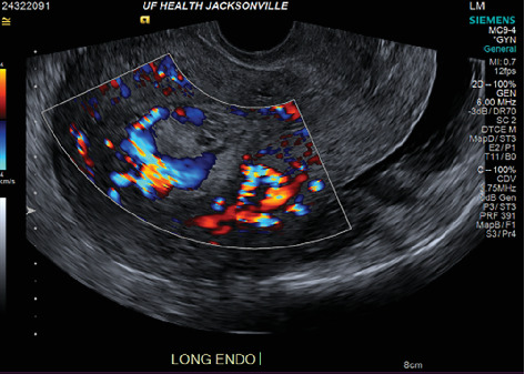
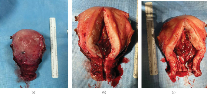
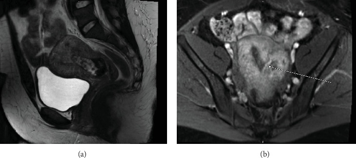
A placental polyp is a retained fragment of placental tissue that can lead to postpartum hemorrhage or become a nidus for infection. Hypervascular placental polyps can pose an increased risk of life-threatening postpartum hemorrhage requiring immediate intervention. Thus, prompt recognition and appropriate management are crucial in preventing maternal morbidity and mortality. Here, we present the case of a 29-year-old patient who had a spontaneous vaginal delivery at 36-week gestation after induction of labor due to pre-eclampsia with severe features. Quantitative blood loss at delivery was 1300 mL, and the patient received uterotonic medications. Due to continued bleeding, she underwent a suction curettage with clots and retained tissue removed from the uterine fundus. The total blood loss was estimated to be 4 L, and the massive transfusion protocol was activated. On postpartum Day 1, she underwent a bilateral uterine artery embolization; however, she developed further heavy vaginal bleeding. A second suction curettage was performed after ultrasound showed hypervascular material in the uterine cavity. The patient was subsequently discharged, but represented on postpartum Day 15 with increased bleeding. Imaging again demonstrated a hypervascular intrauterine polypoid mass. The patient desired definitive management and underwent a minimally invasive total hysterectomy.

 求助内容:
求助内容: 应助结果提醒方式:


