Mehran Rahbar, Ali Sharifi, Javad Hayati Garjan, Mojtaba Sheykhian
{"title":"评估正颌手术患者下颌支和髁突体积的分形值:一项系统回顾和荟萃分析。","authors":"Mehran Rahbar, Ali Sharifi, Javad Hayati Garjan, Mojtaba Sheykhian","doi":"10.1186/s40902-025-00468-7","DOIUrl":null,"url":null,"abstract":"<p><strong>Aim: </strong>The aim of the present study was to evaluate condylar volume and mandibular ramus in patients undergoing orthognathic surgery.</p><p><strong>Method: </strong>Relevant keywords were searched in the international databases Cochrane, Embase, and MEDLINE up to February 2025. Study selection criteria were based on the PICOS strategy; randomized clinical trial studies, cohort studies, cross-sectional studies, case-control studies (study (S)) that examined changes in condylar and ramus position (Outcome (O)) in orthognathic surgery as skeletal treatment (Intervention (I)) for Class III versus Class II (Comparison (C)) in patients who had undergone orthognathic surgery (Population (P)). Data were collected based on study findings from three-dimensional (3D) cephalometric/cone-beam computed tomographic (CBCT)analysis and measurements of condylar angle, volume, and position. The methodological index for non-randomized studies (MINORS) used to determine the quality of the studies. Mean differences were used as an effect size with fixed-effects model and inverse-variance methods of 95% confidence intervals (CI). Meta-analysis was performed using Stata (as of version 17).</p><p><strong>Result: </strong>The mean differences in condylar height between Class II and Class III were 2.19 mm (MD 2.19 mm 95% CI; 1.32 mm, 3.96 mm; p < 0.05). The mean differences in ramus angle between Class II and Class III were - 0.02° (MD - 0.02 95% CI - 0.06, 0.03; p > 0.05).</p><p><strong>Conclusion: </strong>Based on the meta-analysis of the present study, orthognathic surgery did not significantly affect the microstructure of the mandibular ramus in the correction of class III malocclusions. In Class II, the condyle height was significantly reduced after orthognathic surgery, while the condyle width did not change.</p><p><strong>Trial registration: </strong>PROSPERO CRD420251054773.</p>","PeriodicalId":18357,"journal":{"name":"Maxillofacial Plastic and Reconstructive Surgery","volume":"47 1","pages":"14"},"PeriodicalIF":2.8000,"publicationDate":"2025-07-01","publicationTypes":"Journal Article","fieldsOfStudy":null,"isOpenAccess":false,"openAccessPdf":"https://www.ncbi.nlm.nih.gov/pmc/articles/PMC12214064/pdf/","citationCount":"0","resultStr":"{\"title\":\"Evaluating fractal value of mandibular ramus and condylar volume in patients undergoing orthognathic surgery: a systematic review and meta-analysis.\",\"authors\":\"Mehran Rahbar, Ali Sharifi, Javad Hayati Garjan, Mojtaba Sheykhian\",\"doi\":\"10.1186/s40902-025-00468-7\",\"DOIUrl\":null,\"url\":null,\"abstract\":\"<p><strong>Aim: </strong>The aim of the present study was to evaluate condylar volume and mandibular ramus in patients undergoing orthognathic surgery.</p><p><strong>Method: </strong>Relevant keywords were searched in the international databases Cochrane, Embase, and MEDLINE up to February 2025. Study selection criteria were based on the PICOS strategy; randomized clinical trial studies, cohort studies, cross-sectional studies, case-control studies (study (S)) that examined changes in condylar and ramus position (Outcome (O)) in orthognathic surgery as skeletal treatment (Intervention (I)) for Class III versus Class II (Comparison (C)) in patients who had undergone orthognathic surgery (Population (P)). Data were collected based on study findings from three-dimensional (3D) cephalometric/cone-beam computed tomographic (CBCT)analysis and measurements of condylar angle, volume, and position. The methodological index for non-randomized studies (MINORS) used to determine the quality of the studies. Mean differences were used as an effect size with fixed-effects model and inverse-variance methods of 95% confidence intervals (CI). Meta-analysis was performed using Stata (as of version 17).</p><p><strong>Result: </strong>The mean differences in condylar height between Class II and Class III were 2.19 mm (MD 2.19 mm 95% CI; 1.32 mm, 3.96 mm; p < 0.05). The mean differences in ramus angle between Class II and Class III were - 0.02° (MD - 0.02 95% CI - 0.06, 0.03; p > 0.05).</p><p><strong>Conclusion: </strong>Based on the meta-analysis of the present study, orthognathic surgery did not significantly affect the microstructure of the mandibular ramus in the correction of class III malocclusions. In Class II, the condyle height was significantly reduced after orthognathic surgery, while the condyle width did not change.</p><p><strong>Trial registration: </strong>PROSPERO CRD420251054773.</p>\",\"PeriodicalId\":18357,\"journal\":{\"name\":\"Maxillofacial Plastic and Reconstructive Surgery\",\"volume\":\"47 1\",\"pages\":\"14\"},\"PeriodicalIF\":2.8000,\"publicationDate\":\"2025-07-01\",\"publicationTypes\":\"Journal Article\",\"fieldsOfStudy\":null,\"isOpenAccess\":false,\"openAccessPdf\":\"https://www.ncbi.nlm.nih.gov/pmc/articles/PMC12214064/pdf/\",\"citationCount\":\"0\",\"resultStr\":null,\"platform\":\"Semanticscholar\",\"paperid\":null,\"PeriodicalName\":\"Maxillofacial Plastic and Reconstructive Surgery\",\"FirstCategoryId\":\"1085\",\"ListUrlMain\":\"https://doi.org/10.1186/s40902-025-00468-7\",\"RegionNum\":0,\"RegionCategory\":null,\"ArticlePicture\":[],\"TitleCN\":null,\"AbstractTextCN\":null,\"PMCID\":null,\"EPubDate\":\"\",\"PubModel\":\"\",\"JCR\":\"Q2\",\"JCRName\":\"DENTISTRY, ORAL SURGERY & MEDICINE\",\"Score\":null,\"Total\":0}","platform":"Semanticscholar","paperid":null,"PeriodicalName":"Maxillofacial Plastic and Reconstructive Surgery","FirstCategoryId":"1085","ListUrlMain":"https://doi.org/10.1186/s40902-025-00468-7","RegionNum":0,"RegionCategory":null,"ArticlePicture":[],"TitleCN":null,"AbstractTextCN":null,"PMCID":null,"EPubDate":"","PubModel":"","JCR":"Q2","JCRName":"DENTISTRY, ORAL SURGERY & MEDICINE","Score":null,"Total":0}
引用次数: 0
摘要
目的:本研究的目的是评估正颌手术患者的髁突体积和下颌支。方法:检索截至2025年2月的国际数据库Cochrane、Embase和MEDLINE的相关关键词。研究选择标准基于PICOS策略;随机临床试验研究,队列研究,横断面研究,病例对照研究(研究(S)),检查正颌手术作为骨骼治疗(干预(I)) III类与II类(比较(C))接受正颌手术的患者(人群(P))的髁突和支位置的变化(结果(O))。数据收集基于三维(3D)头位测量/锥束计算机断层扫描(CBCT)分析和测量髁角、体积和位置的研究结果。用于确定研究质量的非随机研究(minor)的方法学指数。采用固定效应模型和95%置信区间(CI)的反方差方法,采用平均差异作为效应量。meta分析使用Stata进行(截至版本17)。结果:II类与III类髁突高度的平均差值为2.19 mm (MD为2.19 mm, 95% CI;1.32 mm, 3.96 mm;p 0.05)。结论:基于本研究的meta分析,矫正III类错颌时,正颌手术对下颌支的微结构无明显影响。II类患者,正颌手术后髁突高度明显降低,而髁突宽度没有变化。试验注册:PROSPERO CRD420251054773。
Evaluating fractal value of mandibular ramus and condylar volume in patients undergoing orthognathic surgery: a systematic review and meta-analysis.
Aim: The aim of the present study was to evaluate condylar volume and mandibular ramus in patients undergoing orthognathic surgery.
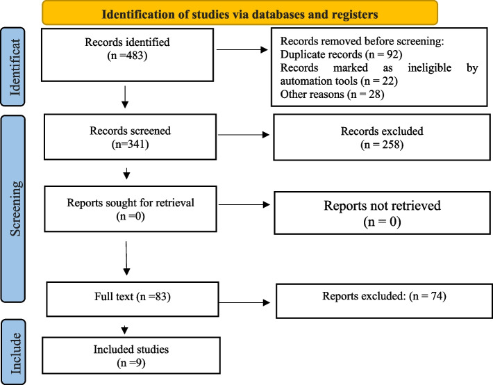
Method: Relevant keywords were searched in the international databases Cochrane, Embase, and MEDLINE up to February 2025. Study selection criteria were based on the PICOS strategy; randomized clinical trial studies, cohort studies, cross-sectional studies, case-control studies (study (S)) that examined changes in condylar and ramus position (Outcome (O)) in orthognathic surgery as skeletal treatment (Intervention (I)) for Class III versus Class II (Comparison (C)) in patients who had undergone orthognathic surgery (Population (P)). Data were collected based on study findings from three-dimensional (3D) cephalometric/cone-beam computed tomographic (CBCT)analysis and measurements of condylar angle, volume, and position. The methodological index for non-randomized studies (MINORS) used to determine the quality of the studies. Mean differences were used as an effect size with fixed-effects model and inverse-variance methods of 95% confidence intervals (CI). Meta-analysis was performed using Stata (as of version 17).
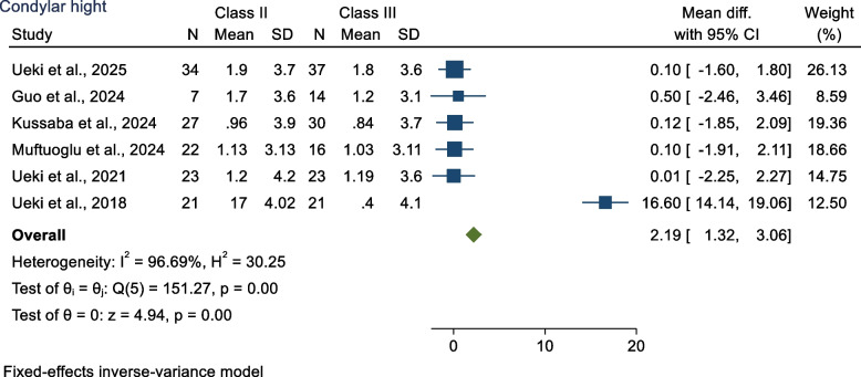
Result: The mean differences in condylar height between Class II and Class III were 2.19 mm (MD 2.19 mm 95% CI; 1.32 mm, 3.96 mm; p < 0.05). The mean differences in ramus angle between Class II and Class III were - 0.02° (MD - 0.02 95% CI - 0.06, 0.03; p > 0.05).
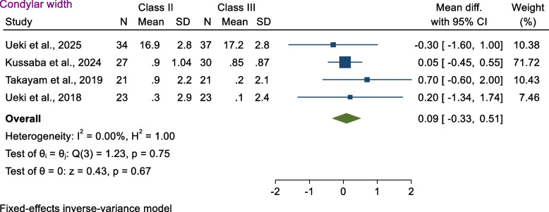
Conclusion: Based on the meta-analysis of the present study, orthognathic surgery did not significantly affect the microstructure of the mandibular ramus in the correction of class III malocclusions. In Class II, the condyle height was significantly reduced after orthognathic surgery, while the condyle width did not change.

 求助内容:
求助内容: 应助结果提醒方式:


