{"title":"利帕舒地尔通过抗氧化机制对视网膜神经节细胞的神经保护作用。","authors":"Reiko Yamagishi-Kimura, Megumi Honjo, Makoto Aihara","doi":"10.1007/s10384-025-01243-x","DOIUrl":null,"url":null,"abstract":"<p><strong>Purpose: </strong>To evaluate the neuroprotective effect of ripasudil, a rho-kinase inhibitor that is a commercially available glaucoma medication that lowers intraocular pressure. We explored the effects of ripasudil on retinal damage via oxidative stress (OS) in primary rat retinal ganglion cell (RGC) cultures and NMDA-induced retinal damage in mice.</p><p><strong>Study design: </strong>Experimental investigation METHODS: Primary rat RGCs were isolated via a 2-step immunopanning method and cultured under normal cultivation conditions for 72 h and for a further 24 h in antioxidant-free medium for OS. We measured the number of living RGCs by use of calcein-AM and calpain activity via calpain immunoreactivity assays. Furthermore, we evaluated the effects of ripasudil via RGC counting in retinal flat-mounts from Thy1-CFP mice, retinal thickness via optical coherence tomography, and reduced glutathione levels via GSSG/GSH assays in NMDA-induced retinal damage.</p><p><strong>Results: </strong>The living RGC counts of normal, OS, 0.1, 1, 10, and 100 uM ripasudil under OS were 236.0 ± 21.6, 155.0 ± 13.2, 155.9 ± 17.1, 158.9 ± 12.0, 184.8 ± 26.9, and 201.1 ± 24.8 cells, respectively. 10 or 100 uM ripasudil significantly inhibited the OS-induced RGC reduction (P < 0.05 or 0.01). Furthermore, the enhanced calpain activity induced by OS was suppressed by 100 uM ripasudil (P < 0.05). In an in vivo study, the RGC counts in the NMDA-treated group were lower than those of the non-NMDA-treated group. NMDA-induced RGC loss was significantly suppressed by ripasudil (P < 0.01). Retinal thinning after 3 weeks of NMDA injection was also inhibited by ripasudil (P < 0.01 or 0.05). Furthermore, NMDA increased the glutathione level, whereas ripasudil suppressed it (P < 0.05).</p><p><strong>Conclusions: </strong>Ripasudil may have neuroprotective effects via an antioxidative mechanism, which could be useful as an intraocular pressure-independent additive.</p>","PeriodicalId":14563,"journal":{"name":"Japanese Journal of Ophthalmology","volume":" ","pages":"823-832"},"PeriodicalIF":1.9000,"publicationDate":"2025-09-01","publicationTypes":"Journal Article","fieldsOfStudy":null,"isOpenAccess":false,"openAccessPdf":"https://www.ncbi.nlm.nih.gov/pmc/articles/PMC12391152/pdf/","citationCount":"0","resultStr":"{\"title\":\"Neuroprotective effect of ripasudil on retinal ganglion cells via an antioxidative mechanism.\",\"authors\":\"Reiko Yamagishi-Kimura, Megumi Honjo, Makoto Aihara\",\"doi\":\"10.1007/s10384-025-01243-x\",\"DOIUrl\":null,\"url\":null,\"abstract\":\"<p><strong>Purpose: </strong>To evaluate the neuroprotective effect of ripasudil, a rho-kinase inhibitor that is a commercially available glaucoma medication that lowers intraocular pressure. We explored the effects of ripasudil on retinal damage via oxidative stress (OS) in primary rat retinal ganglion cell (RGC) cultures and NMDA-induced retinal damage in mice.</p><p><strong>Study design: </strong>Experimental investigation METHODS: Primary rat RGCs were isolated via a 2-step immunopanning method and cultured under normal cultivation conditions for 72 h and for a further 24 h in antioxidant-free medium for OS. We measured the number of living RGCs by use of calcein-AM and calpain activity via calpain immunoreactivity assays. Furthermore, we evaluated the effects of ripasudil via RGC counting in retinal flat-mounts from Thy1-CFP mice, retinal thickness via optical coherence tomography, and reduced glutathione levels via GSSG/GSH assays in NMDA-induced retinal damage.</p><p><strong>Results: </strong>The living RGC counts of normal, OS, 0.1, 1, 10, and 100 uM ripasudil under OS were 236.0 ± 21.6, 155.0 ± 13.2, 155.9 ± 17.1, 158.9 ± 12.0, 184.8 ± 26.9, and 201.1 ± 24.8 cells, respectively. 10 or 100 uM ripasudil significantly inhibited the OS-induced RGC reduction (P < 0.05 or 0.01). Furthermore, the enhanced calpain activity induced by OS was suppressed by 100 uM ripasudil (P < 0.05). In an in vivo study, the RGC counts in the NMDA-treated group were lower than those of the non-NMDA-treated group. NMDA-induced RGC loss was significantly suppressed by ripasudil (P < 0.01). Retinal thinning after 3 weeks of NMDA injection was also inhibited by ripasudil (P < 0.01 or 0.05). Furthermore, NMDA increased the glutathione level, whereas ripasudil suppressed it (P < 0.05).</p><p><strong>Conclusions: </strong>Ripasudil may have neuroprotective effects via an antioxidative mechanism, which could be useful as an intraocular pressure-independent additive.</p>\",\"PeriodicalId\":14563,\"journal\":{\"name\":\"Japanese Journal of Ophthalmology\",\"volume\":\" \",\"pages\":\"823-832\"},\"PeriodicalIF\":1.9000,\"publicationDate\":\"2025-09-01\",\"publicationTypes\":\"Journal Article\",\"fieldsOfStudy\":null,\"isOpenAccess\":false,\"openAccessPdf\":\"https://www.ncbi.nlm.nih.gov/pmc/articles/PMC12391152/pdf/\",\"citationCount\":\"0\",\"resultStr\":null,\"platform\":\"Semanticscholar\",\"paperid\":null,\"PeriodicalName\":\"Japanese Journal of Ophthalmology\",\"FirstCategoryId\":\"3\",\"ListUrlMain\":\"https://doi.org/10.1007/s10384-025-01243-x\",\"RegionNum\":3,\"RegionCategory\":\"医学\",\"ArticlePicture\":[],\"TitleCN\":null,\"AbstractTextCN\":null,\"PMCID\":null,\"EPubDate\":\"2025/7/2 0:00:00\",\"PubModel\":\"Epub\",\"JCR\":\"Q2\",\"JCRName\":\"OPHTHALMOLOGY\",\"Score\":null,\"Total\":0}","platform":"Semanticscholar","paperid":null,"PeriodicalName":"Japanese Journal of Ophthalmology","FirstCategoryId":"3","ListUrlMain":"https://doi.org/10.1007/s10384-025-01243-x","RegionNum":3,"RegionCategory":"医学","ArticlePicture":[],"TitleCN":null,"AbstractTextCN":null,"PMCID":null,"EPubDate":"2025/7/2 0:00:00","PubModel":"Epub","JCR":"Q2","JCRName":"OPHTHALMOLOGY","Score":null,"Total":0}
Neuroprotective effect of ripasudil on retinal ganglion cells via an antioxidative mechanism.
Purpose: To evaluate the neuroprotective effect of ripasudil, a rho-kinase inhibitor that is a commercially available glaucoma medication that lowers intraocular pressure. We explored the effects of ripasudil on retinal damage via oxidative stress (OS) in primary rat retinal ganglion cell (RGC) cultures and NMDA-induced retinal damage in mice.
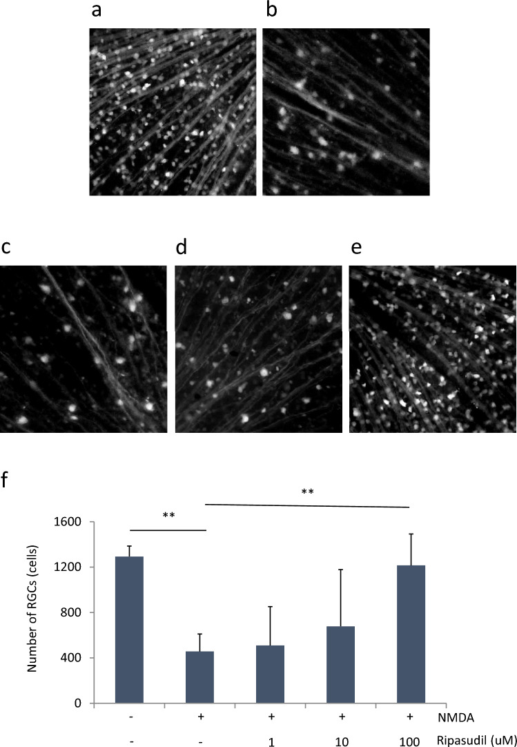
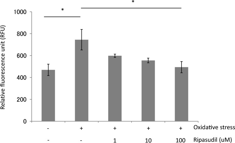
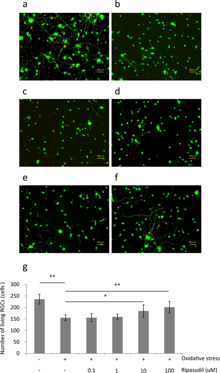
Study design: Experimental investigation METHODS: Primary rat RGCs were isolated via a 2-step immunopanning method and cultured under normal cultivation conditions for 72 h and for a further 24 h in antioxidant-free medium for OS. We measured the number of living RGCs by use of calcein-AM and calpain activity via calpain immunoreactivity assays. Furthermore, we evaluated the effects of ripasudil via RGC counting in retinal flat-mounts from Thy1-CFP mice, retinal thickness via optical coherence tomography, and reduced glutathione levels via GSSG/GSH assays in NMDA-induced retinal damage.
Results: The living RGC counts of normal, OS, 0.1, 1, 10, and 100 uM ripasudil under OS were 236.0 ± 21.6, 155.0 ± 13.2, 155.9 ± 17.1, 158.9 ± 12.0, 184.8 ± 26.9, and 201.1 ± 24.8 cells, respectively. 10 or 100 uM ripasudil significantly inhibited the OS-induced RGC reduction (P < 0.05 or 0.01). Furthermore, the enhanced calpain activity induced by OS was suppressed by 100 uM ripasudil (P < 0.05). In an in vivo study, the RGC counts in the NMDA-treated group were lower than those of the non-NMDA-treated group. NMDA-induced RGC loss was significantly suppressed by ripasudil (P < 0.01). Retinal thinning after 3 weeks of NMDA injection was also inhibited by ripasudil (P < 0.01 or 0.05). Furthermore, NMDA increased the glutathione level, whereas ripasudil suppressed it (P < 0.05).
Conclusions: Ripasudil may have neuroprotective effects via an antioxidative mechanism, which could be useful as an intraocular pressure-independent additive.
期刊介绍:
The Japanese Journal of Ophthalmology (JJO) was inaugurated in 1957 as a quarterly journal published in English by the Ophthalmology Department of the University of Tokyo, with the aim of disseminating the achievements of Japanese ophthalmologists worldwide. JJO remains the only Japanese ophthalmology journal published in English. In 1997, the Japanese Ophthalmological Society assumed the responsibility for publishing the Japanese Journal of Ophthalmology as its official English-language publication.
Currently the journal is published bimonthly and accepts papers from authors worldwide. JJO has become an international interdisciplinary forum for the publication of basic science and clinical research papers.

 求助内容:
求助内容: 应助结果提醒方式:


