Martina Čančarević, Vjekoslav Radeljić, Matias Trbušić, Zdravko Babić, Ivan Zeljković, Nikola Kos
{"title":"无st抬高合并冠状动脉穿孔及心包填塞的急性心肌梗死经皮小斜支介入治疗。","authors":"Martina Čančarević, Vjekoslav Radeljić, Matias Trbušić, Zdravko Babić, Ivan Zeljković, Nikola Kos","doi":"10.20471/acc.2024.63.s1.9","DOIUrl":null,"url":null,"abstract":"<p><strong>Introduction: </strong>Small coronary artery disease is more common in elderly patients, smokers, patients with diabetes and chronic kidney disease. Percutaneous interventions on small coronary arteries are associated with an increased risk of complications (perforation, dissection and restenosis). Coronary artery perforation treatment includes cover stents and coil placement.</p><p><strong>Case report: </strong>A 73-year-old patient, without comorbidities, was hospitalized for acute non ST-elevation myocardial infarction. Coronary angiography showed subocclusion of the first diagonal branch (culprit lesion) while the other epicardial coronary arteries were without stenosis. Multiple predilatations of the target vessel were performed, and as it was a vessel with a diameter of less than 2 mm, no stent was placed. The final angiogram showed normal flow and good morphological result. Half an hour after the procedure, cardiac tamponade and cardiorespiratory arrest developed. Emergency pericardiocentesis was performed and after the return of spontaneous circulation, emergency recoronarography was performed. It showed perforation of the diagonal branch with contrast extravasation. Coronary coil was applied proximal to the perforation site. Perforation repair and hemodynamic stabilization were achieved.</p><p><strong>Conclusion: </strong>Coronary artery perforation is a life-threatening complication of percutaneous coronary intervention. The risk of perforation is higher in the case of small coronary arteries; it can be presented by delayed cardiac tamponade, which requires increased supervision of the patient.</p>","PeriodicalId":7072,"journal":{"name":"Acta clinica Croatica","volume":"63 Suppl1","pages":"47-53"},"PeriodicalIF":0.8000,"publicationDate":"2024-03-01","publicationTypes":"Journal Article","fieldsOfStudy":null,"isOpenAccess":false,"openAccessPdf":"https://www.ncbi.nlm.nih.gov/pmc/articles/PMC12207842/pdf/","citationCount":"0","resultStr":"{\"title\":\"PERCUTANEOUS CORONARY INTERVENTION OF THE SMALL DIAGONAL BRANCH IN ACUTE MYOCARDIAL INFARCTION WITHOUT ST ELEVATION COMPLICATED BY CORONARY ARTERY PERFORATION AND CARDIAC TAMPONADE.\",\"authors\":\"Martina Čančarević, Vjekoslav Radeljić, Matias Trbušić, Zdravko Babić, Ivan Zeljković, Nikola Kos\",\"doi\":\"10.20471/acc.2024.63.s1.9\",\"DOIUrl\":null,\"url\":null,\"abstract\":\"<p><strong>Introduction: </strong>Small coronary artery disease is more common in elderly patients, smokers, patients with diabetes and chronic kidney disease. Percutaneous interventions on small coronary arteries are associated with an increased risk of complications (perforation, dissection and restenosis). Coronary artery perforation treatment includes cover stents and coil placement.</p><p><strong>Case report: </strong>A 73-year-old patient, without comorbidities, was hospitalized for acute non ST-elevation myocardial infarction. Coronary angiography showed subocclusion of the first diagonal branch (culprit lesion) while the other epicardial coronary arteries were without stenosis. Multiple predilatations of the target vessel were performed, and as it was a vessel with a diameter of less than 2 mm, no stent was placed. The final angiogram showed normal flow and good morphological result. Half an hour after the procedure, cardiac tamponade and cardiorespiratory arrest developed. Emergency pericardiocentesis was performed and after the return of spontaneous circulation, emergency recoronarography was performed. It showed perforation of the diagonal branch with contrast extravasation. Coronary coil was applied proximal to the perforation site. Perforation repair and hemodynamic stabilization were achieved.</p><p><strong>Conclusion: </strong>Coronary artery perforation is a life-threatening complication of percutaneous coronary intervention. The risk of perforation is higher in the case of small coronary arteries; it can be presented by delayed cardiac tamponade, which requires increased supervision of the patient.</p>\",\"PeriodicalId\":7072,\"journal\":{\"name\":\"Acta clinica Croatica\",\"volume\":\"63 Suppl1\",\"pages\":\"47-53\"},\"PeriodicalIF\":0.8000,\"publicationDate\":\"2024-03-01\",\"publicationTypes\":\"Journal Article\",\"fieldsOfStudy\":null,\"isOpenAccess\":false,\"openAccessPdf\":\"https://www.ncbi.nlm.nih.gov/pmc/articles/PMC12207842/pdf/\",\"citationCount\":\"0\",\"resultStr\":null,\"platform\":\"Semanticscholar\",\"paperid\":null,\"PeriodicalName\":\"Acta clinica Croatica\",\"FirstCategoryId\":\"3\",\"ListUrlMain\":\"https://doi.org/10.20471/acc.2024.63.s1.9\",\"RegionNum\":4,\"RegionCategory\":\"医学\",\"ArticlePicture\":[],\"TitleCN\":null,\"AbstractTextCN\":null,\"PMCID\":null,\"EPubDate\":\"\",\"PubModel\":\"\",\"JCR\":\"Q3\",\"JCRName\":\"MEDICINE, GENERAL & INTERNAL\",\"Score\":null,\"Total\":0}","platform":"Semanticscholar","paperid":null,"PeriodicalName":"Acta clinica Croatica","FirstCategoryId":"3","ListUrlMain":"https://doi.org/10.20471/acc.2024.63.s1.9","RegionNum":4,"RegionCategory":"医学","ArticlePicture":[],"TitleCN":null,"AbstractTextCN":null,"PMCID":null,"EPubDate":"","PubModel":"","JCR":"Q3","JCRName":"MEDICINE, GENERAL & INTERNAL","Score":null,"Total":0}
PERCUTANEOUS CORONARY INTERVENTION OF THE SMALL DIAGONAL BRANCH IN ACUTE MYOCARDIAL INFARCTION WITHOUT ST ELEVATION COMPLICATED BY CORONARY ARTERY PERFORATION AND CARDIAC TAMPONADE.
Introduction: Small coronary artery disease is more common in elderly patients, smokers, patients with diabetes and chronic kidney disease. Percutaneous interventions on small coronary arteries are associated with an increased risk of complications (perforation, dissection and restenosis). Coronary artery perforation treatment includes cover stents and coil placement.
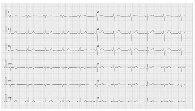
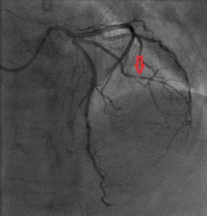
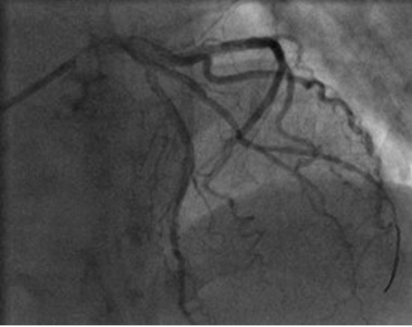
Case report: A 73-year-old patient, without comorbidities, was hospitalized for acute non ST-elevation myocardial infarction. Coronary angiography showed subocclusion of the first diagonal branch (culprit lesion) while the other epicardial coronary arteries were without stenosis. Multiple predilatations of the target vessel were performed, and as it was a vessel with a diameter of less than 2 mm, no stent was placed. The final angiogram showed normal flow and good morphological result. Half an hour after the procedure, cardiac tamponade and cardiorespiratory arrest developed. Emergency pericardiocentesis was performed and after the return of spontaneous circulation, emergency recoronarography was performed. It showed perforation of the diagonal branch with contrast extravasation. Coronary coil was applied proximal to the perforation site. Perforation repair and hemodynamic stabilization were achieved.
Conclusion: Coronary artery perforation is a life-threatening complication of percutaneous coronary intervention. The risk of perforation is higher in the case of small coronary arteries; it can be presented by delayed cardiac tamponade, which requires increased supervision of the patient.
期刊介绍:
Acta Clinica Croatica is a peer reviewed general medical journal that publishes original articles that advance and improve medical science and practice and that serve the purpose of transfer of original and valuable information to journal readers. Acta Clinica Croatica is published in English four times a year.
 求助内容:
求助内容: 应助结果提醒方式:


